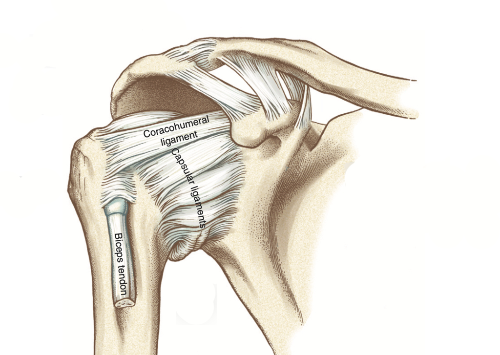
Acromioclavicular joint (AC)
By : Yaser MohammedLocation and Articulation
Acromioclavicular joint (AC): Is a joint in the upper limp, it works as a connection area between the arm and the chest. Is one of four joints that comprises the Shoulder Complex.
This acromioclavicular joint is formed from the articulation of two bones. The first is the Scapula articulate at its acromion process (The scapula has 3 processes: acromion, spine, and Coracoid process), While the second bone is the clavicle(collar bone) at its lateral end (part), which describes the reason for the naming.
Note
Note: Shoulder complex joints are sternoclavicular (SC), acromioclavicular (AC), and scapulothoracic joints, and glenohumeral joint.This acromioclavicular joint is formed from the articulation of two bones. The first is the Scapula articulate at its acromion process (The scapula has 3 processes: acromion, spine, and Coracoid process), While the second bone is the clavicle(collar bone) at its lateral end (part), which describes the reason for the naming.
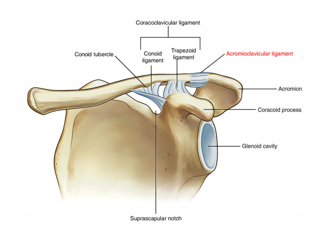
Acromioclavicular joint articulation
Joint Type and Its Movement
The joint is a plane synovial joint; multiaxial (has three degrees of freedom), which under normal physiological conditions allows gliding movement (function).
Having the clavicle as a bone in common, the movements within the acromioclavicular and sternoclavicular joints are closely related to each other. And as it attaches the scapula to the thorax, it allows an additional range of motion to the scapula and assists in arm movement such as shoulder abduction and flexion.
The three degrees of freedom of acromioclavicular joints are:
1) Protraction – retraction
Protraction: The protraction of the scapula on the AC joint is primarily produced by the serratus anterior and pectoralis minor muscles.
Retraction: While retraction of scapula at this joint occurs due to contraction of the horizontal and ascending parts of the trapezius muscle, and rhomboid minor and major muscles
2) Elevation - depression (total range of motion 15°)
3) Lateral rotation - medial rotation (total range of motion 30°)
Having the clavicle as a bone in common, the movements within the acromioclavicular and sternoclavicular joints are closely related to each other. And as it attaches the scapula to the thorax, it allows an additional range of motion to the scapula and assists in arm movement such as shoulder abduction and flexion.
The three degrees of freedom of acromioclavicular joints are:
1) Protraction – retraction
Protraction: The protraction of the scapula on the AC joint is primarily produced by the serratus anterior and pectoralis minor muscles.
Retraction: While retraction of scapula at this joint occurs due to contraction of the horizontal and ascending parts of the trapezius muscle, and rhomboid minor and major muscles
2) Elevation - depression (total range of motion 15°)
3) Lateral rotation - medial rotation (total range of motion 30°)
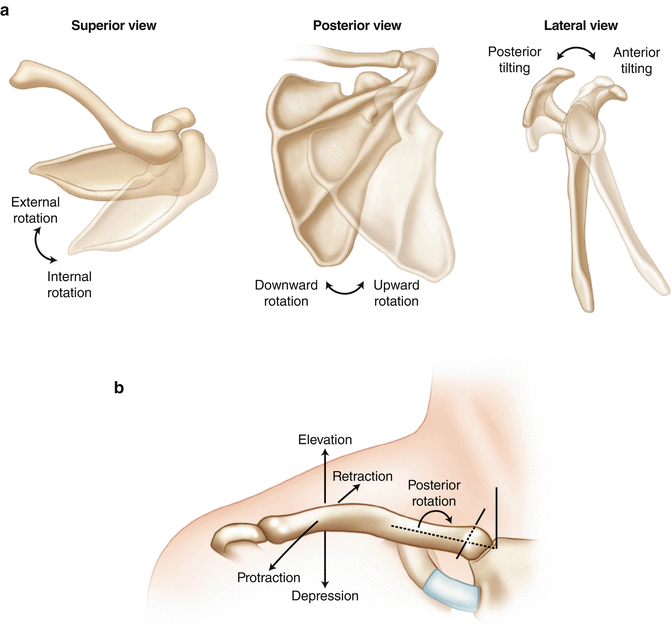

Acromioclavicular joint Motion
Blood Supply
The Blood supply of this joint is the Thoracoacromial artery , and the Suprascapular artery , The first is a branch of the axillary artery, while the second stems from the subclavian artery via the thyrocervical trunk.
Nerve Innervation
Consistent with Hilton’s law (joints are supplied by articular
branches of the nerves supplying the muscles that act on
the joint), The acromioclavicular joint is innervated by the lateral pectoral and suprascapular nerves . The lateral pectoral nerve is a branch of the lateral cord of brachial plexus, while the suprascapular nerves originate from the superior trunk of the plexus.
branches of the nerves supplying the muscles that act on
the joint), The acromioclavicular joint is innervated by the lateral pectoral and suprascapular nerves . The lateral pectoral nerve is a branch of the lateral cord of brachial plexus, while the suprascapular nerves originate from the superior trunk of the plexus.
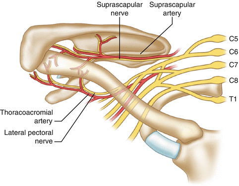
Illustration of the neurovascular anatomy that supplies the AC joint. The suprascapular nerve and lateral pectoral nerve provide the innervation while the suprascapular artery and thoracoacromial artery provide the blood supply.
Capsule and Ligaments
The AC capsule (fibrous capsule)and ligaments surrounding the joint work together to provide stability and keep the clavicle in contact with the acromion process of the scapula.
The AC Joint has a thin capsule lined, it's weak and strengthed inferiorly and superiorly by capsular ligaments.
The ligaments of this joint are:
1) Coracoclavicular Ligaments: Is a strong pair of bands that unite the coracoid process of the scapula to the clavicle and run from the coracoid process of the scapula to the underside clavicle.
2) Conoid ligament: is an inverted triangle (cone), which has its apex inferiorly it extends from the posterior surface of the coracoid process to the conoid tubercle of the clavicle.
3) Trapezoid ligament: Extends from the superior surface of the coracoid process to the trapezoid line on the inferior surface of the clavicle.
4) Acromioclavicular ligament: A fibrous band extending from the acromion to the clavicle that strengthens the AC joint superiorly found within the joint capsule, is composed of two ligaments superior and inferior acromioclavicular ligament.
The AC Joint has a thin capsule lined, it's weak and strengthed inferiorly and superiorly by capsular ligaments.
The ligaments of this joint are:
1) Coracoclavicular Ligaments: Is a strong pair of bands that unite the coracoid process of the scapula to the clavicle and run from the coracoid process of the scapula to the underside clavicle.
2) Conoid ligament: is an inverted triangle (cone), which has its apex inferiorly it extends from the posterior surface of the coracoid process to the conoid tubercle of the clavicle.
3) Trapezoid ligament: Extends from the superior surface of the coracoid process to the trapezoid line on the inferior surface of the clavicle.
4) Acromioclavicular ligament: A fibrous band extending from the acromion to the clavicle that strengthens the AC joint superiorly found within the joint capsule, is composed of two ligaments superior and inferior acromioclavicular ligament.
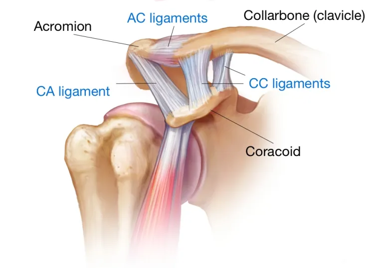
The acromioclavicular (AC) and coracoclavicular (CC) ligaments
Acromioclavicular Joint Dislocation
Acute dislocation of the acromioclavicular joint is a common injury in athletes and is characterized by painful impairment of shoulder function and elevation of the lateral clavicle.
The AC joint is the only bony connection of your shoulder blade and is essential for maintaining normal dynamics in the shoulder.
AC joint stability depends mainly on three structures: AC joint capsule, the coracoclavicular ligaments, and the delto-trapezial fascia.
The AC joint is the only bony connection of your shoulder blade and is essential for maintaining normal dynamics in the shoulder.
AC joint stability depends mainly on three structures: AC joint capsule, the coracoclavicular ligaments, and the delto-trapezial fascia.
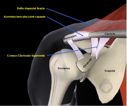
The AC joint stability factors
a fall on the tip of the shoulder can cause an injury in the joint and become unstable and painful. This injury is most common in men and normally occurs between ages (18 – 45) and likely occurs in collision sports, such as rugby, or other sports that can cause a fall in the shoulder.
AC joint dislocation can be classified into six grades , the degree of the injury does not directly represent the degree of symptoms, This grade is determined by x-rays and clinical examination, which can help us to determine which structure is damaged.
Grades 1 and 2: No prominence in the shoulder, AC joint will be stable, the joint capsule has been injured, but the essential coracoclavicular ligaments will be intact.
Grade 3: a prominence found on the shoulder, On the x-ray clavicle, has moved out of its socket. The coracoclavicular ligaments have been injured, but the delto-trapezial fascia probably remains intact.
Grade 4: similar to Grade 3 but in this case, the clavicle is unstable posteriorly, which causes the clavicle to penetrate the trapezius muscle behind the joint which causes problems.
Grade 5: In this case, the overlying delto-trapezial fascia has also ruptured and the clavicle has displaced its original position.
Grade 6: is a rare injury pattern where the clavicle comes beneath the coracoid process.
AC joint dislocation can be classified into six grades , the degree of the injury does not directly represent the degree of symptoms, This grade is determined by x-rays and clinical examination, which can help us to determine which structure is damaged.
Grades 1 and 2: No prominence in the shoulder, AC joint will be stable, the joint capsule has been injured, but the essential coracoclavicular ligaments will be intact.
Grade 3: a prominence found on the shoulder, On the x-ray clavicle, has moved out of its socket. The coracoclavicular ligaments have been injured, but the delto-trapezial fascia probably remains intact.
Grade 4: similar to Grade 3 but in this case, the clavicle is unstable posteriorly, which causes the clavicle to penetrate the trapezius muscle behind the joint which causes problems.
Grade 5: In this case, the overlying delto-trapezial fascia has also ruptured and the clavicle has displaced its original position.
Grade 6: is a rare injury pattern where the clavicle comes beneath the coracoid process.
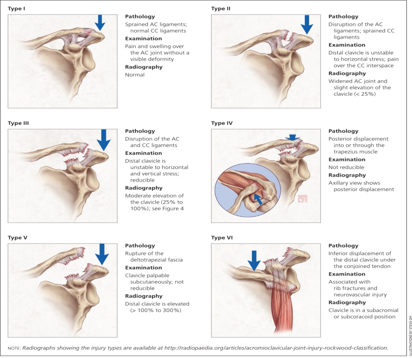
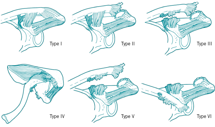
The Six grades of acromioclavicular joint dislocation
References
1) Moore K. L., Dalley, A. F., & Agur, A. M. R. (2014)- Clinically Oriented Anatomy 7th Edition (796-813)
2) Netter, F. (2019). Atlas of Human Anatomy (7th ed.). Philadelphia, PA: Saunders.
3) Acromioclavicular (AC) joint , Jana Vasković MD , Kenhub
https://www.kenhub.com/en/library/anatomy/acromioclavicular-ac-joint
4) The Acromioclavicular Joint, Peter J. Millett2 , Musculoskeletal Key
https://musculoskeletalkey.com/the-acromioclavicular-joint/
5) The Acromioclavicular Joint, Rosamond Nicolson and Jaspreet Johal, Teachme Anatomy
https://teachmeanatomy.info/upper-limb/joints/acromioclavicular/
6) Acromioclavicular joint dislocation, Windsor upper limb
https://www.windsorupperlimb.com/conditions/shoulder-conditions/common-shoulder-fractures/acromioclavicular-joint-dislocation
2) Netter, F. (2019). Atlas of Human Anatomy (7th ed.). Philadelphia, PA: Saunders.
3) Acromioclavicular (AC) joint , Jana Vasković MD , Kenhub
https://www.kenhub.com/en/library/anatomy/acromioclavicular-ac-joint
4) The Acromioclavicular Joint, Peter J. Millett2 , Musculoskeletal Key
https://musculoskeletalkey.com/the-acromioclavicular-joint/
5) The Acromioclavicular Joint, Rosamond Nicolson and Jaspreet Johal, Teachme Anatomy
https://teachmeanatomy.info/upper-limb/joints/acromioclavicular/
6) Acromioclavicular joint dislocation, Windsor upper limb
https://www.windsorupperlimb.com/conditions/shoulder-conditions/common-shoulder-fractures/acromioclavicular-joint-dislocation
References of images
Cover Image : Shoulder ligaments Diagram | Created by
ae2621 | Quizlet https://o.quizlet.com/qz2KEEyxD-TMxDrMF-S8Mw_b.png
fig 2: Acromioclavicular joint motion, Musculoskeletal Key
https://musculoskeletalkey.com/wp-content/uploads/2016/10/A312451_1_En_7_Fig11_HTML.gif
fig 3: Movements of the acromioclavicular joint (diagram), Kenhub
https://www.kenhub.com/thumbor/qde0HZmhl7o51Da0-TBRYrMSHeA=/fit-in/800x1600/filters:watermark(/images/logo_url.png,-10,-10,0):background_color(FFFFFF):format(jpeg)/images/library/13128/ACJ_axes.png
fig 4: Illustration of the neurovascular anatomy that supplies the AC joint. The suprascapular nerve and lateral pectoral nerve provide the innervation while the suprascapular artery and thoracoacromial artery provide the blood supply. Musculoskeletal Key
https://musculoskeletalkey.com/wp-content/uploads/2016/10/A312451_1_En_7_Fig10_HTML.gif
fig 5 : The acromioclavicular (AC) and coracoclavicular (CC) ligaments, Shoulderelbow.
https://i0.wp.com/shoulderelbow.org/wp-content/uploads/2016/09/acromioclavicular-and-coracoclavicular-ligaments.jpg?resize=748%2C538&ssl=1
fig 6: AC joint stability factors, Windsor
http://www.windsorupperlimb.com/data/images/Acromio%20joint%202.png
fig 7: The Six grades of acromioclavicular joint dislocation, Aspetar Sports Medicine journals.
https://www.aspetar.com/journal/upload/images/thump/2014127154057.png
ae2621 | Quizlet https://o.quizlet.com/qz2KEEyxD-TMxDrMF-S8Mw_b.png
fig 2: Acromioclavicular joint motion, Musculoskeletal Key
https://musculoskeletalkey.com/wp-content/uploads/2016/10/A312451_1_En_7_Fig11_HTML.gif
fig 3: Movements of the acromioclavicular joint (diagram), Kenhub
https://www.kenhub.com/thumbor/qde0HZmhl7o51Da0-TBRYrMSHeA=/fit-in/800x1600/filters:watermark(/images/logo_url.png,-10,-10,0):background_color(FFFFFF):format(jpeg)/images/library/13128/ACJ_axes.png
fig 4: Illustration of the neurovascular anatomy that supplies the AC joint. The suprascapular nerve and lateral pectoral nerve provide the innervation while the suprascapular artery and thoracoacromial artery provide the blood supply. Musculoskeletal Key
https://musculoskeletalkey.com/wp-content/uploads/2016/10/A312451_1_En_7_Fig10_HTML.gif
fig 5 : The acromioclavicular (AC) and coracoclavicular (CC) ligaments, Shoulderelbow.
https://i0.wp.com/shoulderelbow.org/wp-content/uploads/2016/09/acromioclavicular-and-coracoclavicular-ligaments.jpg?resize=748%2C538&ssl=1
fig 6: AC joint stability factors, Windsor
http://www.windsorupperlimb.com/data/images/Acromio%20joint%202.png
fig 7: The Six grades of acromioclavicular joint dislocation, Aspetar Sports Medicine journals.
https://www.aspetar.com/journal/upload/images/thump/2014127154057.png