
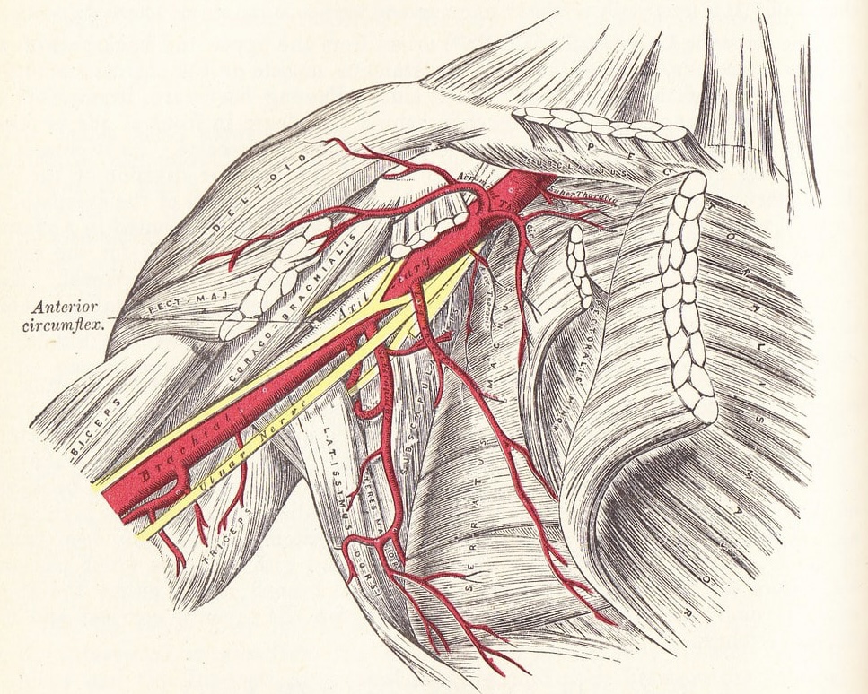
Axillary Artery
By : Ali JalalDefinition
Axillary artery, Arteria axillaris ,is the largest muscular artery that travels through the axilla.

Course
It begins as a continuation of subclavian artery at the lateral border of the first rib.
It runs behind the pectoralis minor muscle into the arm,then it becomes the brachial artery at lower border of teres major muscle.
Throughout its course, the artery is closely related to the cords of the brachial plexus and their branches and is enclosed with them in a connective tissue sheath called the axillary sheath .
It runs behind the pectoralis minor muscle into the arm,then it becomes the brachial artery at lower border of teres major muscle.
Throughout its course, the artery is closely related to the cords of the brachial plexus and their branches and is enclosed with them in a connective tissue sheath called the axillary sheath .

Parts of the axillary artery
Axillary artery is divided into three parts according to the pectoralis minor muscle which is in front of it.
The parts :
1) The first part: which originates from the lateral border of 1st rib and ends at rhe upper border of pectoralis minor muscle.
2) The second part : which lies behind the pectoralis minor muscle.
3) The third part : which begins at the lower border of the pectoralis minor muscle to the lower border of the teres major muscle.
The parts :
1) The first part: which originates from the lateral border of 1st rib and ends at rhe upper border of pectoralis minor muscle.
2) The second part : which lies behind the pectoralis minor muscle.
3) The third part : which begins at the lower border of the pectoralis minor muscle to the lower border of the teres major muscle.
Branches of the axillary artery
A)From the first part:
-the highest thoracic artery : which is small and runs along the upper border of pectoralis minor muscle.
B)From the second part:
-the thoracoacromial artery :which divides into terminal branches:
1-pectoral
2-clavicular
3-deltoid
4-acromial
deep to clavicular head of the pectoralis major muscle.
-the lateral thoracic artery :runs along the lower border of the pectoralis minor muscle.
C)From the third part:
-the subscapular artery :which runs along the lower border of the subscapularis muscle on the posterior axillary wall. it soon terminates by dividing into the circumflex scapular and thoracoacromial arteries.
-the anterior and posterior circumference humeral arteries :these arteries wind around the front and the back of the surgical neck of the humerus respectively.
-the highest thoracic artery : which is small and runs along the upper border of pectoralis minor muscle.
B)From the second part:
-the thoracoacromial artery :which divides into terminal branches:
1-pectoral
2-clavicular
3-deltoid
4-acromial
deep to clavicular head of the pectoralis major muscle.
-the lateral thoracic artery :runs along the lower border of the pectoralis minor muscle.
C)From the third part:
-the subscapular artery :which runs along the lower border of the subscapularis muscle on the posterior axillary wall. it soon terminates by dividing into the circumflex scapular and thoracoacromial arteries.
-the anterior and posterior circumference humeral arteries :these arteries wind around the front and the back of the surgical neck of the humerus respectively.

Clinical Note
Critical Stenosis of axillary artery
Focal axillary artery stenosis or occlusion is a rare-occurrence case that is most commonly encountered after severe trauma involving the shoulder joint or proximal humerus.
Other etiologies that can lead to axillary artery injury or pathology include different vasculitides, radiation arteritis, crutch-injury, and peripheral arterial disease.
It could be treated by percutaneous angioplasty and stenting .
Focal axillary artery stenosis or occlusion is a rare-occurrence case that is most commonly encountered after severe trauma involving the shoulder joint or proximal humerus.
Other etiologies that can lead to axillary artery injury or pathology include different vasculitides, radiation arteritis, crutch-injury, and peripheral arterial disease.
It could be treated by percutaneous angioplasty and stenting .

References
1. Keith L. Moore , Arthur F. Dalley A. M. R. Agur; Moore clinically oriented anatomy 7thedition;pp.93,99,699,715-718
2. Axillary artery;Lorenzo Crumbie MBBS,BSc;Kenhub; https://www.kenhub.com/en/library/anatomy/the-axillary-artery
3. Anatomy, Shoulder and Upper Limb, Axillary Artery;Robert Thiel, Akul Munjal, Daniel T. Daly;National Library of Medicine; https://www.ncbi.nlm.nih.gov/books/NBK482174/
4. Critical stenosis of axillary artery treated with percutaneous angioplasty and stenting: a case report and review of theliterature; JonathanKim,BS,YoussefElias,MD, PhD;Sciencedirect;https://www.sciencedirect.com/science/article/pii/S1930043322003946
2. Axillary artery;Lorenzo Crumbie MBBS,BSc;Kenhub; https://www.kenhub.com/en/library/anatomy/the-axillary-artery
3. Anatomy, Shoulder and Upper Limb, Axillary Artery;Robert Thiel, Akul Munjal, Daniel T. Daly;National Library of Medicine; https://www.ncbi.nlm.nih.gov/books/NBK482174/
4. Critical stenosis of axillary artery treated with percutaneous angioplasty and stenting: a case report and review of theliterature; JonathanKim,BS,YoussefElias,MD, PhD;Sciencedirect;https://www.sciencedirect.com/science/article/pii/S1930043322003946