

Glenohumeral Joint (GH) (Shoulder Joint)
By : Yaser MohammedLocation and Articulation
Glenohumeral Joint (GH): Is a joint in the upper limp, it works as a connection area between the body and the two arms. Is one of four joints that comprises the Shoulder Complex.
The glenohumeral joint is formed from the articulation of the convex head (proximal end) of the humerus and the concave glenoid fossa(glenoid cavity) of the scapula(lateral part of the scapula). Both articular surfaces are covered with hyaline cartilage .
Note
Note: Shoulder complex joints are sternoclavicular (SC), acromioclavicular (AC), scapulothoracic joints, and glenohumeral joints.The glenohumeral joint is formed from the articulation of the convex head (proximal end) of the humerus and the concave glenoid fossa(glenoid cavity) of the scapula(lateral part of the scapula). Both articular surfaces are covered with hyaline cartilage .

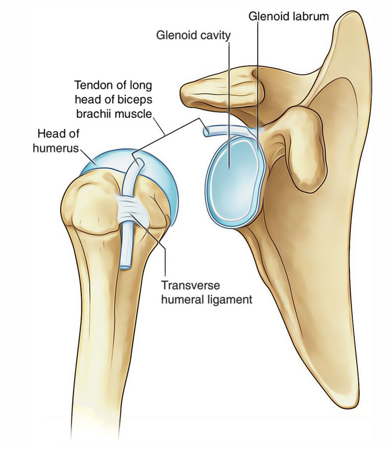
Glenohumeral Joint Articulation
Joint Type and Its Movement
The joint is a true ball-and-socket synovial joint , multiaxial (move at multiple planes), and the GH joint is the most mobile joint in the body, This freedom results from the laxity of its joint capsule and the large size of the humeral head compared with the small size of the glenoid cavity.
however, this high mobility affects joint stability, Making GH joint the most commonly dislocated diarthrodial joint.
The GH joint work with the joint of the shoulder complex to make the movement at the shoulder.
The glenohumeral joint allows movements around three axe:
1) Flexion (110°) - extension (60°): shoulder flexion is when you move your arms forward anywhere from a resting position by your sides to straight above your head. An extension is when you move your arms and stick them out behind you.
flexion is acted by pectoralis major, deltoid, coracobrachialis and long head of biceps brachii muscles. While the extension is acted mainly by Deltoid and assisted by latissimus dorsi, teres major, pectoralis major, and long head of triceps brachii muscles.
2) Abduction (120°) - adduction (0°): The abduction is the upward lateral movement of humerus out to the side, away from the body. While adduction is the downward movement of the humerus medially toward the body.
Abduction is acted by Supraspinatus and deltoid muscles. While adduction by coracobrachialis, pectoralis major, latissimus dorsi, and teres major muscles.
3) Internal rotation (90°) - external rotation (90°): Internal shoulder rotation involves rotating your upper arm toward the front side of your trunk. External rotation involves rotating it away from the front side of your trunk.
internal rotation is acted mainly by Subscapularis and is also assisted by teres major, latissimus dorsi, pectoralis major, and deltoid muscles. While external rotation is mainly by infraspinatus, Teres minor, and deltoid muscles.
however, this high mobility affects joint stability, Making GH joint the most commonly dislocated diarthrodial joint.
The GH joint work with the joint of the shoulder complex to make the movement at the shoulder.
The glenohumeral joint allows movements around three axe:
1) Flexion (110°) - extension (60°): shoulder flexion is when you move your arms forward anywhere from a resting position by your sides to straight above your head. An extension is when you move your arms and stick them out behind you.
flexion is acted by pectoralis major, deltoid, coracobrachialis and long head of biceps brachii muscles. While the extension is acted mainly by Deltoid and assisted by latissimus dorsi, teres major, pectoralis major, and long head of triceps brachii muscles.
2) Abduction (120°) - adduction (0°): The abduction is the upward lateral movement of humerus out to the side, away from the body. While adduction is the downward movement of the humerus medially toward the body.
Abduction is acted by Supraspinatus and deltoid muscles. While adduction by coracobrachialis, pectoralis major, latissimus dorsi, and teres major muscles.
3) Internal rotation (90°) - external rotation (90°): Internal shoulder rotation involves rotating your upper arm toward the front side of your trunk. External rotation involves rotating it away from the front side of your trunk.
internal rotation is acted mainly by Subscapularis and is also assisted by teres major, latissimus dorsi, pectoralis major, and deltoid muscles. While external rotation is mainly by infraspinatus, Teres minor, and deltoid muscles.

The Movements of GH Joint

Basic Shoulder Movements
Note: Rotator Cuffs
Rotator cuff muscles are four muscles that form a musculotendinous unit around the shoulder joint. Which are the supraspinatus, infraspinatus, teres minor , and subscapularis muscles (to memorize them we can be called them SITS ), These muscles provide individuals with the ability to lift their arms and reach overhead.
.png)

Muscles Around The GH joint
Blood Supply
The Blood supply of this joint is the anterior and posterior circumflex humeral, suprascapular, and circumflex scapular arteries. [see fig 3]
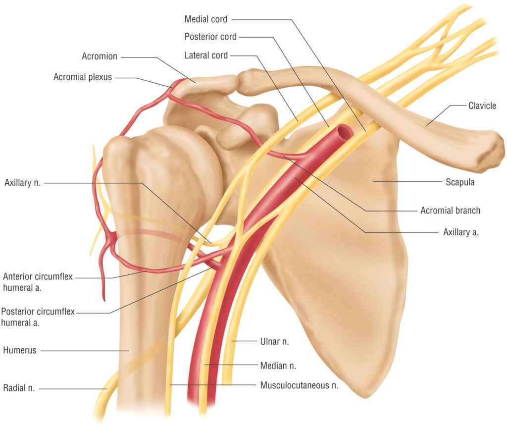
Blood Supply of Glenohumeral Joint
Nerve Innervation
While the Nerve innervation is the subscapular nerve (C5-C6), which is a branch of the posterior cord of the brachial plexus.
The joint capsule is also supplied by many sources:
• Suprascapular nerve (superior and posterior parts)
• Axillary nerve (anteroinferior part),
• Lateral pectoral nerve (anterosuperior part of the capsule).
The joint capsule is also supplied by many sources:
• Suprascapular nerve (superior and posterior parts)
• Axillary nerve (anteroinferior part),
• Lateral pectoral nerve (anterosuperior part of the capsule).

Nerve Innervation of Glenohumeral Joint
Joint Capsule
The shoulder joint is encircled by a loose fibrous capsule . It extends from the scapula to the humerus, enclosing the joint on all sides. The internal surface of the capsule is lined by a synovial membrane.
This joint capsule has two openings:
1) An opening between the tubercles of the humerus for passage of the tendon of the long head of the biceps brachii.
2) Between the superior and middle glenohumeral ligaments, via which the subscapular bursa communicates with the glenohumeral joint cavity.
This joint capsule has two openings:
1) An opening between the tubercles of the humerus for passage of the tendon of the long head of the biceps brachii.
2) Between the superior and middle glenohumeral ligaments, via which the subscapular bursa communicates with the glenohumeral joint cavity.

Anterior view of the capsule of the right glenohumeral joint
Joints Ligaments
The GH joint has several ligaments that cover it to stabilize the joint and resist dislocations, these are the glenohumeral, coracohumeral, and transverse humeral ligaments.
transverse humeral and Glenohumeral ligaments are called capsular ligaments, while coracohumeral is called accessory ligaments.
Transverse humeral ligament: this ligament extends horizontally between the two tubercles of the humerus. It covers the intertubercular sulcus and the long head tendon of the biceps brachii muscle.
Coracohumeral ligament: this ligament extends between the coracoid process of the scapula to the two tubercles of the humerus and the transverse humeral ligament covering.
Glenohumeral ligaments: It is a three ligaments superior, middle, and inferior that support the joint from the anteroinferior side.
Superior glenohumeral ligament: extends from the supraglenoid tubercle of the scapula to the proximal aspect of the lesser tubercle of the humerus.
Middle glenohumeral ligament: extends from the anterior glenoid margin of the scapula to the lesser tubercle of the humerus.
Inferior glenohumeral ligament: extends between the glenoid labrum and the inferomedial part of the humeral neck, it's a sling-like ligament, it divides into anterior and posterior bands and is the strongest ligament of the GH ligaments.
transverse humeral and Glenohumeral ligaments are called capsular ligaments, while coracohumeral is called accessory ligaments.
Transverse humeral ligament: this ligament extends horizontally between the two tubercles of the humerus. It covers the intertubercular sulcus and the long head tendon of the biceps brachii muscle.
Coracohumeral ligament: this ligament extends between the coracoid process of the scapula to the two tubercles of the humerus and the transverse humeral ligament covering.
Glenohumeral ligaments: It is a three ligaments superior, middle, and inferior that support the joint from the anteroinferior side.
Superior glenohumeral ligament: extends from the supraglenoid tubercle of the scapula to the proximal aspect of the lesser tubercle of the humerus.
Middle glenohumeral ligament: extends from the anterior glenoid margin of the scapula to the lesser tubercle of the humerus.
Inferior glenohumeral ligament: extends between the glenoid labrum and the inferomedial part of the humeral neck, it's a sling-like ligament, it divides into anterior and posterior bands and is the strongest ligament of the GH ligaments.

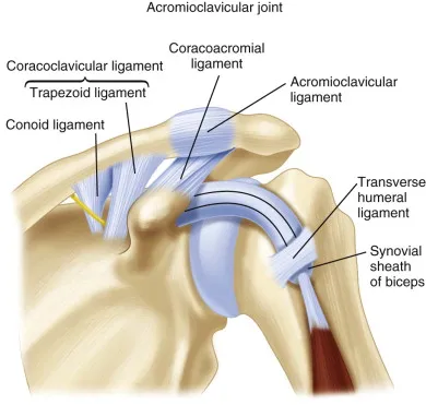
The ligaments of the shoulder joint. The transverse humeral ligament is not shown in this diagram
References
1) Moore K. L., Dalley, A. F., & Agur, A. M. R. (2014)- Clinically Oriented Anatomy 7th Edition (796-800 plates).
2) Netter, F. (2019). Atlas of Human Anatomy (7th ed.). Philadelphia, PA: Saunders.
3) Glenohumeral joint ,Nicola McLaren MSc , Kenhub.
https://www.kenhub.com/en/library/anatomy/the-shoulder-joint
4) Glenohumeral Joint , Physiopedia
https://www.physio-pedia.com/Glenohumeral_Joint
5) The Shoulder Joint , Oliver Jones , Teachme Anatomy.
https://teachmeanatomy.info/upper-limb/joints/shoulder/
2) Netter, F. (2019). Atlas of Human Anatomy (7th ed.). Philadelphia, PA: Saunders.
3) Glenohumeral joint ,Nicola McLaren MSc , Kenhub.
https://www.kenhub.com/en/library/anatomy/the-shoulder-joint
4) Glenohumeral Joint , Physiopedia
https://www.physio-pedia.com/Glenohumeral_Joint
5) The Shoulder Joint , Oliver Jones , Teachme Anatomy.
https://teachmeanatomy.info/upper-limb/joints/shoulder/
References of images
Cover photo: Anterior view of the left shoulder and acromioclavicular joints, and proper ligaments of the scapula. Henry Gray (1918) Anatomy of the Human Body Bartleby.com: Gray's Anatomy, Plate 326
fig 1: Glenohumeral joint articulation, Illustration by Maya Chastain, Healthline
https://i0.wp.com/post.healthline.com/wp-content/uploads/2022/05/2248093-Glenohumeral-Osteoarthritis-Causes-Symptoms-and-Treatment-1296x1085-Body.jpg?w=1155&h=2278
fig 2: Shoulder Joint (Glenohumeral Joint) – Earth's Lab.
fig 4: Basic shoulder movements, moushuspilates.com.
https://moushuspilates.com/wp-content/uploads/2020/11/Shoulder-movements-768x329.png
https://www.earthslab.com/wp-content/uploads/2018/08/shoulder-joint.jpg
fig 6: Muscles on the dorsum of the scapula, and the Triceps brachii.
Henry Gray (1918) Anatomy of the Human Body Bartleby.com: Gray's Anatomy, plate 412.
fig 7: Shoulder Joint | Medical Junction.
https://medical-junction.com/wp-content/uploads/2022/03/blood-and-nerve-supply-shoulder-joint.jpg
fig 8: Schematic diagram of sensory innervation of the shoulder joint (A, anterior view ; B posterior view). ReasearchGate.
https://www.researchgate.net/profile/Pierre-Laumonerie/publication/343174815/figure/fig1/AS:920131693707264@1596387899578/Schematic-diagram-of-sensory-innervation-of-the-shoulder-joint-A-anterior-view-B.png
fig 9: Henry Gray (1918) Anatomy of the Human Body Bartleby.com: Gray's Anatomy, Plate 327
fig 10: The ligaments of the shoulder joint. The transverse humeral ligament is not shown on this diagram , Teachme Anatomy.
https://teachmeanatomy.info/wp-content/uploads/The-Major-Ligaments-of-the-Shoulder-Joint.jpg
fig 11: An anterior view of the shoulder demonstrating the coracoclavicular ligaments, acromioclavicular joint, coracoacromial ligament, and transverse humeral ligament.
Musculoskeletal Key. https://i2.wp.com/musculoskeletalkey.com/wp-content/uploads/2019/02/f042-003-9781455743766.jpg?w=960
fig 1: Glenohumeral joint articulation, Illustration by Maya Chastain, Healthline
https://i0.wp.com/post.healthline.com/wp-content/uploads/2022/05/2248093-Glenohumeral-Osteoarthritis-Causes-Symptoms-and-Treatment-1296x1085-Body.jpg?w=1155&h=2278
fig 2: Shoulder Joint (Glenohumeral Joint) – Earth's Lab.
fig 4: Basic shoulder movements, moushuspilates.com.
https://moushuspilates.com/wp-content/uploads/2020/11/Shoulder-movements-768x329.png
https://www.earthslab.com/wp-content/uploads/2018/08/shoulder-joint.jpg
fig 6: Muscles on the dorsum of the scapula, and the Triceps brachii.
Henry Gray (1918) Anatomy of the Human Body Bartleby.com: Gray's Anatomy, plate 412.
fig 7: Shoulder Joint | Medical Junction.
https://medical-junction.com/wp-content/uploads/2022/03/blood-and-nerve-supply-shoulder-joint.jpg
fig 8: Schematic diagram of sensory innervation of the shoulder joint (A, anterior view ; B posterior view). ReasearchGate.
https://www.researchgate.net/profile/Pierre-Laumonerie/publication/343174815/figure/fig1/AS:920131693707264@1596387899578/Schematic-diagram-of-sensory-innervation-of-the-shoulder-joint-A-anterior-view-B.png
fig 9: Henry Gray (1918) Anatomy of the Human Body Bartleby.com: Gray's Anatomy, Plate 327
fig 10: The ligaments of the shoulder joint. The transverse humeral ligament is not shown on this diagram , Teachme Anatomy.
https://teachmeanatomy.info/wp-content/uploads/The-Major-Ligaments-of-the-Shoulder-Joint.jpg
fig 11: An anterior view of the shoulder demonstrating the coracoclavicular ligaments, acromioclavicular joint, coracoacromial ligament, and transverse humeral ligament.
Musculoskeletal Key. https://i2.wp.com/musculoskeletalkey.com/wp-content/uploads/2019/02/f042-003-9781455743766.jpg?w=960