
Midpalmar space
By : Sedra MuhammedIntroduction:
It's part of the facial spaces of the hand specifically the palmar space.
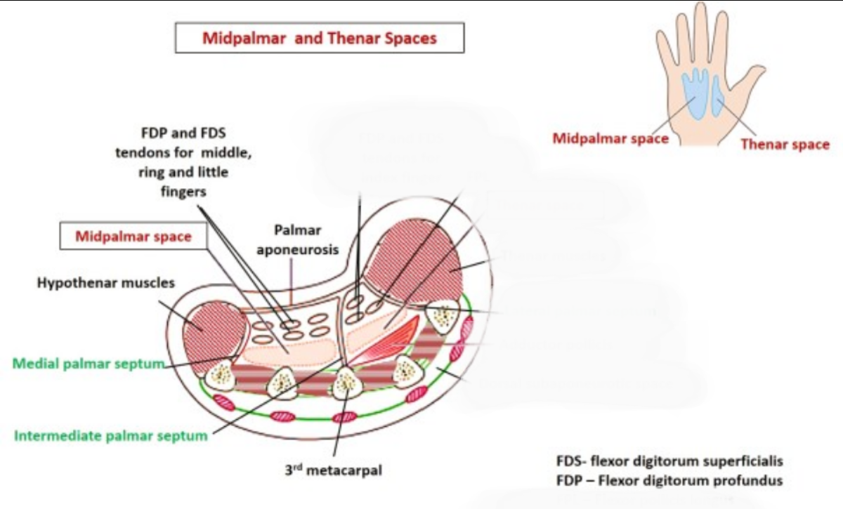
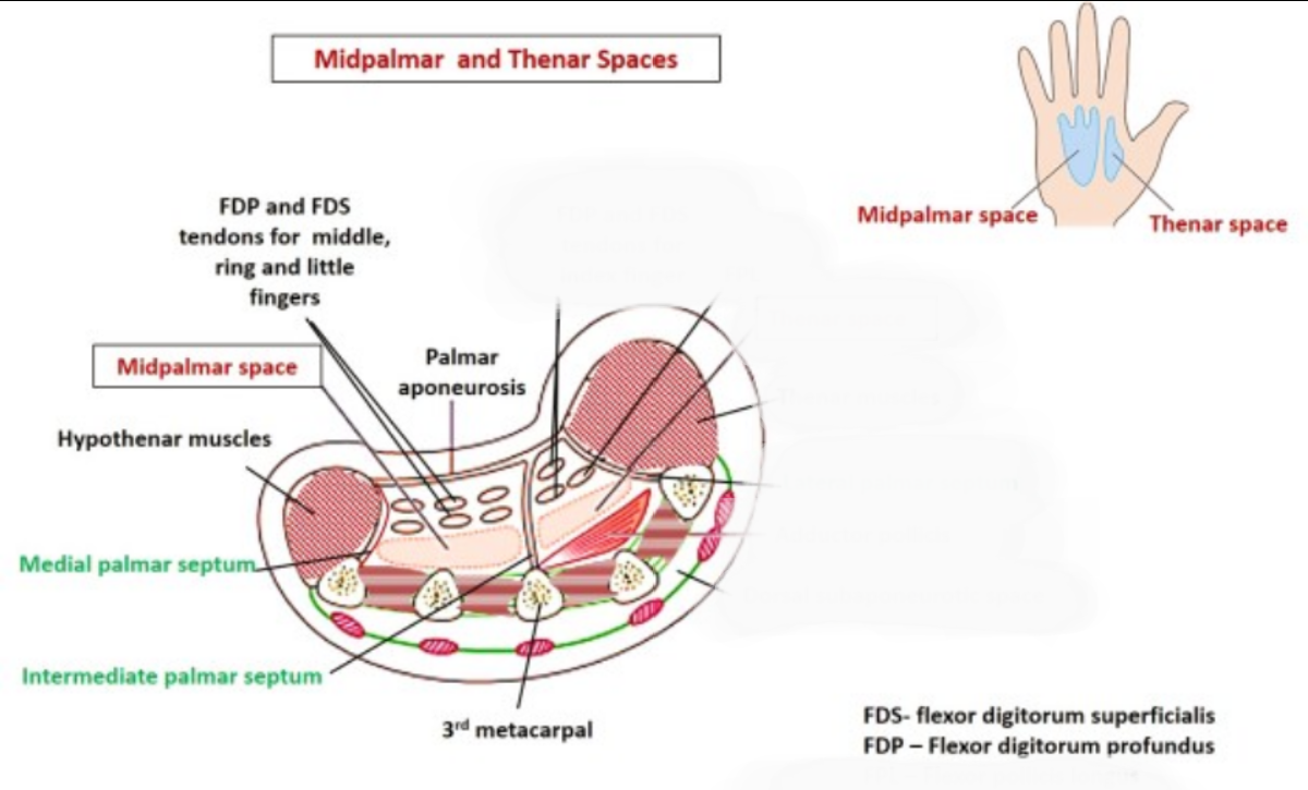
This is a triangular space situated under the medial half of the hollow of the palm.
• Proximally
- it extends up to the distal margin of the flexor retinaculum.
- communicates with the forearm space.
• Distally
- it extends up to the distal palmar crease.
- communicates with the fascial sheaths of the 3rd and 4th lumbrical muscles.
This is a triangular space situated under the medial half of the hollow of the palm.
• Proximally
- it extends up to the distal margin of the flexor retinaculum.
- communicates with the forearm space.
• Distally
- it extends up to the distal palmar crease
Note
distal palmar crease: it’s the farthest crease on your palm among the other two creases, it’s transverse and the nearest to your fingers, It begins close to your little finger and ends at the base of your middle or index finger, or between them.- communicates with the fascial sheaths of the 3rd and 4th lumbrical muscles.
Boundries:
• Anteriorly – palmar aponeurosis:
• Posteriorly – 3rd, 4th and 5th metacarpals the fascia covering the interossei of the 3rd and 4th the spaces, and the medial part of the transverse head of the adductor pollicis
• Medially – medial palmar septum
• Laterally , by the intermediate palmar septum.
• Posteriorly – 3rd, 4th and 5th metacarpals the fascia covering the interossei of the 3rd and 4th the spaces, and the medial part of the transverse head of the adductor pollicis
• Medially – medial palmar septum
• Laterally , by the intermediate palmar septum.
Contents:
• The flexor tendons of third, fourth and fifth fingers
• Second, third and fourth lumbrical muscles;
• Superficial palmar arch
• Digital nerves and vessels of the medial three and half fingers.
• Second, third and fourth lumbrical muscles;
• Superficial palmar arch
• Digital nerves and vessels of the medial three and half fingers.
Clinical notes regarding midpalmar space:
What causes midpalmar space infection?
Infection of the mid palmar space may result from tenosynovitis of the middle and ring fingers, or from a web infection which has spread proximally through the lumbrical canals.
Midpalmar space infections are rare.
What does a midpalmar space infection do?
In a mid-palmar infection the concavity of the palm is usually lost. There is an exquisite tenderness over the infected area which is pallid and sometimes red. Fingers are held rigidly flexed, with decreasing rigidity from the little to the index finger. However, finger rigidity in case of mid-palmar infections is less severe than the rigidity of septic tenosynovitis.
What are the symptoms of a midpalmar space infection?
There is usually a pitting edema in the remaining palm (over the thenar) and at the dorsum of the hand,
while in the case of pus extension along the lumbrical canals, swelling of the web space is also observed.
How to treat a midpalmar space infection?
The midpalmar space is drained and debrided through a palmar incision (transverse, longitudinal or along one of the lumbrical canals) taking care to avoid injury of the neurovascular bundle or contamination of the surrounding tissues (the ulnar bursa in particular).
Infection of the mid palmar space may result from tenosynovitis of the middle and ring fingers, or from a web infection which has spread proximally through the lumbrical canals.
Midpalmar space infections are rare.
What does a midpalmar space infection do?
In a mid-palmar infection the concavity of the palm is usually lost. There is an exquisite tenderness over the infected area which is pallid and sometimes red. Fingers are held rigidly flexed, with decreasing rigidity from the little to the index finger. However, finger rigidity in case of mid-palmar infections is less severe than the rigidity of septic tenosynovitis.
What are the symptoms of a midpalmar space infection?
There is usually a pitting edema
Note
pitting edema: when a swollen part of your body has a dimple (or pit) after you press it for a few seconds.while in the case of pus extension along the lumbrical canals, swelling of the web space is also observed.
How to treat a midpalmar space infection?
The midpalmar space is drained and debrided through a palmar incision (transverse, longitudinal or along one of the lumbrical canals) taking care to avoid injury of the neurovascular bundle or contamination of the surrounding tissues (the ulnar bursa in particular).
References
Bacterial Infections
William D. James MD, in Andrews' Diseases of the Skin, 2020
Hand Infections
Frederick M. Azar MD, in Campbell's Operative Orthopaedics, 2021
Infections of the hand
James Chang MD, in Plastic Surgery: Volume 6: Hand and Upper Extremity, 2018
Incision and Drainage
James R. Roberts MD, FACEP, FAAEM, FACMT, in Roberts and Hedges’ Clinical Procedures in Emergency Medicine and Acute Care, 2019
national library of medicine
closed spaces hand infections: diagnostic and treatment consideration, 2012
Gathier PJ, Schönberger TJ. A man with an infected finger: a case report. J Med Case Rep. 2015
William D. James MD, in Andrews' Diseases of the Skin, 2020
Hand Infections
Frederick M. Azar MD, in Campbell's Operative Orthopaedics, 2021
Infections of the hand
James Chang MD, in Plastic Surgery: Volume 6: Hand and Upper Extremity, 2018
Incision and Drainage
James R. Roberts MD, FACEP, FAAEM, FACMT, in Roberts and Hedges’ Clinical Procedures in Emergency Medicine and Acute Care, 2019
national library of medicine
closed spaces hand infections: diagnostic and treatment consideration, 2012
Gathier PJ, Schönberger TJ. A man with an infected finger: a case report. J Med Case Rep. 2015