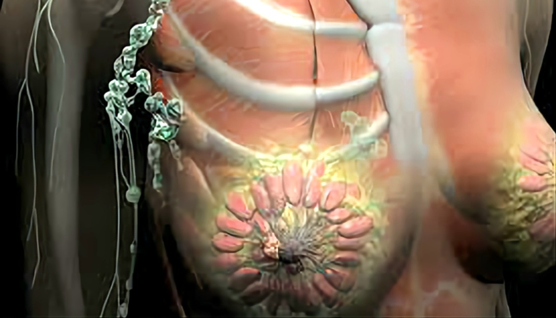
Breast Anatomy
By : Omar M. Subhi Altaie
Breast is a conical-shaped structure that lies in the thoracic region, superficial to the Pectoralis major and Serratus anterior muscles.
The breast has its fundamental functions in nursing and gives newborn babies the right nourishment.
The breast has many severe diseases that may affect the lives of humans, especially cancer in its various types, which makes it important to learn about and explore how to evade the dangers of these diseases that we are all susceptible to.
The Breasts are the most superficial, standing-out structures in the anterior thoracic wall. Breasts consist of glandular tissue (that has specialized to produce milk) as well as fatty tissue; the amount of fat determines the size of the breast.
The fat presents in the male breast is not different from that of subcutaneous tissue somewhere else in the body and the glandular tissue does not develop in any normal case, this structure of the breast in males shares similar characteristics to the breast of those females who have not yet approached the age of puberty.
The fat presents in the male breast is not different from that of subcutaneous tissue somewhere else in the body and the glandular tissue does not develop in any normal case, this structure of the breast in males shares similar characteristics to the breast of those females who have not yet approached the age of puberty.
Female breasts
Under the influence of the ovarian hormone (especially estrogen) when the female approaches the age of puberty (8-15 years old) the breasts assume their hemispherical shape, increasing in the size of the breast is due to the fat deposition as the main reason, but also the glandular development contributes in the size- increasing of the breasts.
The more the female is younger, the more the fat is in small amounts at the expense of glandular development.
The size and shape of the breasts also be determined by genetic, ethnic (or racial), and dietary factors.
• For non-breastfeeding women, the fat surrounding the glandular tissue (that is not completely developed) determines the size of their breasts.
• But, in pregnant women there are several stages that the woman passes through that we are going to discuss as follows:
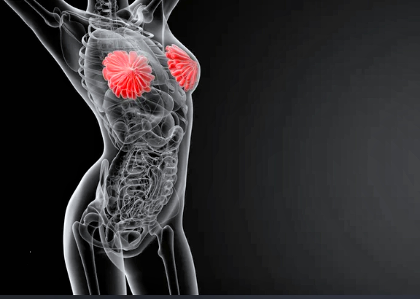
Early
At the beginning of the pregnancy, the duct system gets in rapid increases in length and branching. Blood vascular system of the connective tissue increases; to provide the best nourishment for the glands that have developed.
The alveoli (which are the hollow-like spaces where the milk is secreted ) start to develop.
Nipples enlarge and the areola becomes dark brown due to the melanin pigment deposition in the epidermis.
The areola glands enlarge and become active in the secretary of oily substance that provides lubricant and moisturizer for the nipple and areola.
Late
In the second half of pregnancy the growth process gets slower, even if the breast is still getting to a bigger size; the main reason behind this; because of the swelling or puffiness of the alveoli with the fluid secretion called colostrum .
Colostrum
Is a creamy white to yellow fluid that is secreted before the milk in first-time nursing, it is rich in protein and is high in antibodies and antioxidants to build a newborn immune system and some growth factors that may affect the baby's intestines.
Postweaning
The breasts return to their previous inactive state when the baby has been weaned. The remaining milk is absorbed; the secretory alveoli shrink, and most of them disappear. The interlobular connective tissue thickens. The breasts and the nipples shrink and return nearly to their original size. The pigmentation of the areola gets lighter, but the area never lightens to its original color.
Postmenopause
The breast atrophies (stops functioning as normal) after menopause (when the menstrual cycle stops due to the lower ovarian hormone levels like estrogen and progesterone, normally at the age between 45-55), the alveoli disappear. The amount of adipose tissue may increase or decrease.
Breast position :
The body of the breast lies on the bed of the breast that extends:
Transversely : from the lateral border of the sternum to the midaxillary line.
Vertically : from the second to the sixth rib.
Two-thirds of the bed is formed by pectoral fascia overlying the pectoralis major muscle
The other third of the bed is situated by the fascia that covers the serratus anterior muscle
● between the breast and the fascia there is a space called retromammary space , this space contains amounts of fat that allow the slightly moving of the breast on that fascia.
Axillary tail : formed in the inferior and lateral border of the pectoralis major muscle and pieces the deep fascia in the axillary fossa (armpit)
Nipple and areola
nipples are conical or cylindrical prominences in the centers of the areolae . The nipples have no fat, hair, or sweat glands under them.
The tips of the nipples are fissured (or perforated) with the lactiferous ducts which open independently in the nipple.
The position of the nipple is anterior to the fourth intercostal space for the normal adult female.
The nipples are composed mostly of smooth muscle fibers arranged in circular patterns that compress the lactiferous ducts during lactation and erect the nipples in response to several stimulations, such as when a baby begins to suckle.
The areola pigmented circle in the apex of the breast contains sebaceous glands that secrete an oily substance that provides moisturizer for the nipple and areola.
The color of the areola changes to darker brownish color after the first pregnancy and lasts forever.

Mammary glands
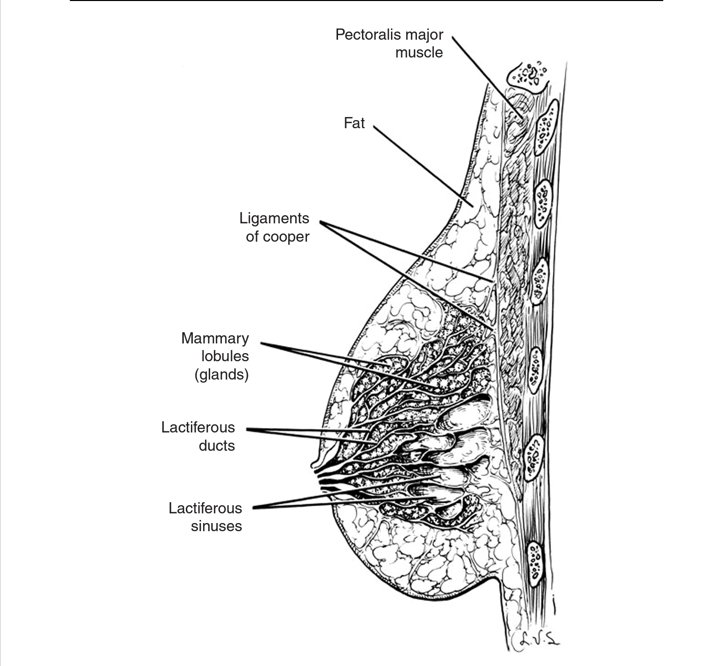
The mammary glands are modified sweat glands, big amounts of these glands are present in the breast in the superficial fascia, and might continue to pierce the deep fascia to the axillary fossa. The upper parts of the mammary glands are well-developed, forming the suspensory ligament (or cooper ligament who described them) this ligament connects the glands to the dermis of the skin overlying it, as well as supports the lobes and lobules of these glands.
The suspensory ligament also maintains the conical shape of the breast.
The Interior of the breast is divided into 15 to 20 components that radiated out from the nipple. The lactiferous ducts continue to become small buds that develop to become 15 to 20 lobules of the mammary glands which together constitute the parenchyma
(The essential and functional substance) of the mammary gland.
Each one of the 15 - 20 lobules drains into a lactiferous duct, which in turn opens independently in the nipple, but just before it opens, it has a dilated ampulla deep to the areola called the lactiferous sinus.
The lactiferous sinus is where the milk droplets accumulate, once the baby starts to suckle compression of the areola and sinus forces the milk to flow out and encourages the baby to keep nursing.
The let-down reflex
Once the baby starts nursing, some small nerves get stimulated, and thus two hormones are released, prolactin which helps to make the milk, and oxytocin forces the milk to be pushed out from where it has been stored. And that when the milk is received in the baby's mouth easily.
Blood supply of the breast
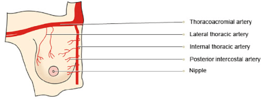
The arterial supply of the breast drives from :
• The second to fourth perforating branches, that pierce the intercostal muscles give the medial mammary branches
And the anterior intercostal branches from that internal thoracic artery originate from the subclavian artery just before it continues as the axillary artery.
• Lateral thoracic artery from the second part of the axillary artery.
• Pectoral branch of Thracoacromial artery originates from the second part of the axillary artery.
• Thoracic aorta gives rise to posterior intercostal arteries in the second third and fourth intercostal spaces that terminate in the breast.
Note : more than half of the blood supply comes from the superior and medial perforators that originate from the medial mammary branches.
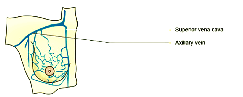
Venous drainage of the breast
The superficial vein follows along the anterior surface of the fascia, these normally drain from the areola and nipple and they sometimes referred to as the venous plexus of Haller
The deep veins probably have the same names as the arteries that they are accompanied by, they mainly drain to the axillary vein and the internal thoracic vein which in turn eventually terminate into the brachiocephalic vein, and to the intercostal veins.
The deep veins probably have the same names as the arteries that they are accompanied by, they mainly drain to the axillary vein and the internal thoracic vein which in turn eventually terminate into the brachiocephalic vein, and to the intercostal veins.
Nerve supply of the breast
Sensory innervation originates from the peripheral nervous system. The fourth to sixth intercostal nerves give rise to the anterior and lateral cutaneous branches, these nerves perforate the pectoralis major muscle and the fascia overlying it to reach the tissue and skin of the breasts, and sensation to the nipples is given by the lateral cutaneous branch of T4.
These Intercostal nerves give sensory fibers to the skin of the breast as well as sympathetic fibers to the blood vessels and smooth muscle in the nipple and skin.
These Intercostal nerves give sensory fibers to the skin of the breast as well as sympathetic fibers to the blood vessels and smooth muscle in the nipple and skin.

Lymph drainage of the breast
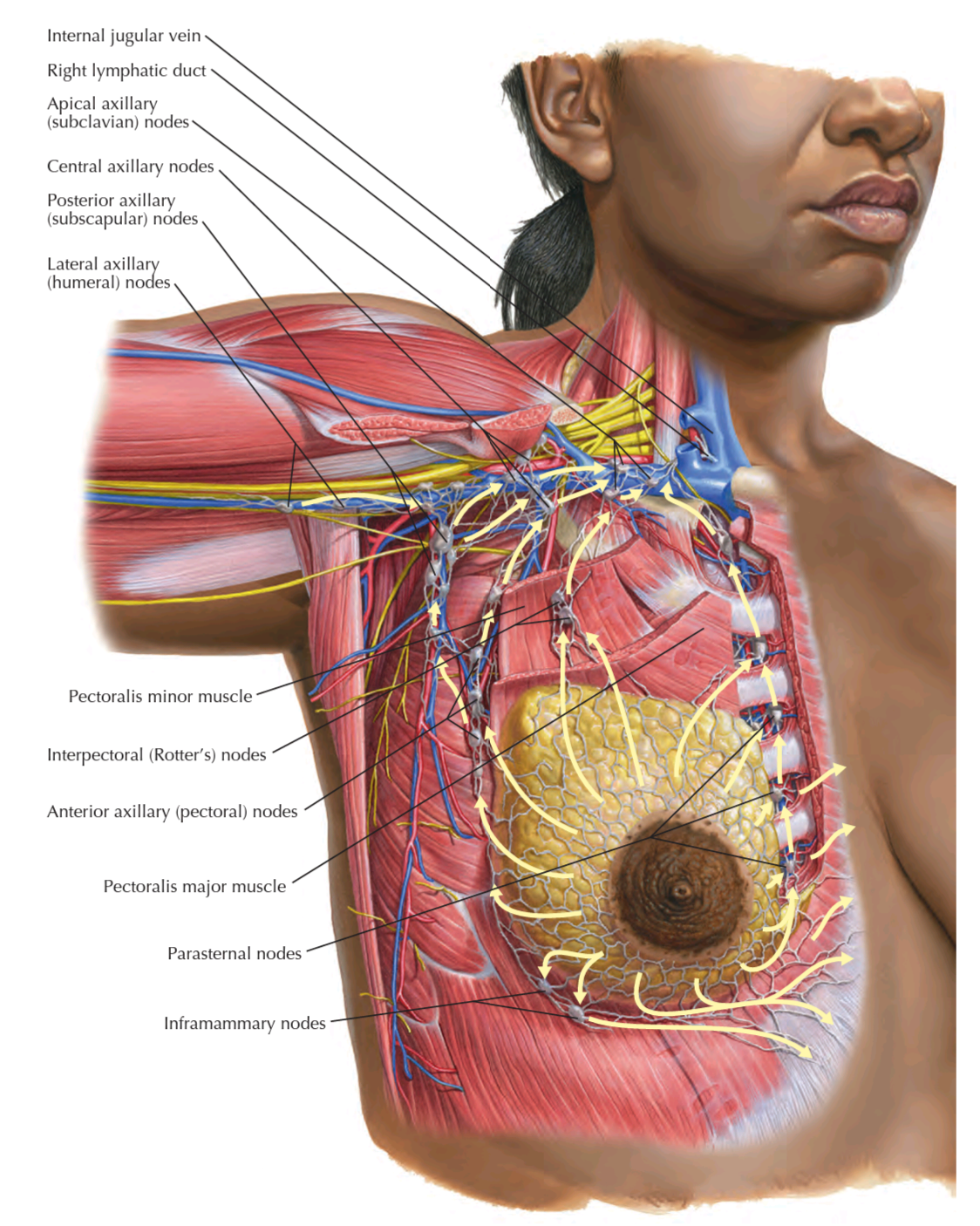
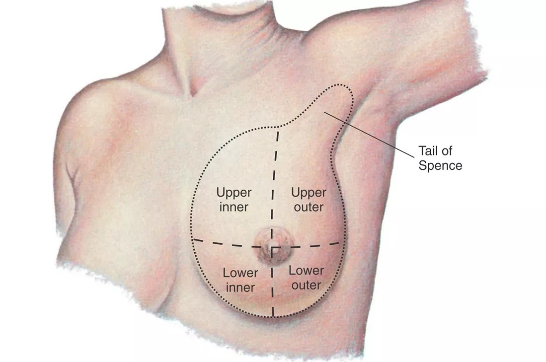
The female breast is divided into four quadrants and a central part, each quadrant drains lymph into a group of lymph nodes near it, and those groups of nodes will eventually drain into :
• groups of nodes of the left breast drain into the thoracic duct which enters the left brachiocephalic vein (at the root of the neck ).
• groups of nodes of the right breast drain into the right lymphatic duct, which opens into the right brachiocephalic vein.
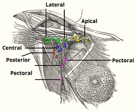
■ Lateral quadrants and central part drain into the axillary groups of lymph nodes
▪︎ lateral quadrant and central part drain into the pectoral group, but the upper quadrant
drain into the apical group.
■ Medial quadrants drain into the parasternal ( or internal thoracic) lymph nodes or may communicate with the opposite breast.
■ lymph from the inferior quadrants drains to abdominal lymph nodes (subdiaphragmatic inferior phrenic lymph nodes).
■ few lymph vessels follow the posterior intercostal arteries to drain into the posterior intercostal nodes
Note
Lymph is a clear or pale-white fluid made of: • White blood cells, especially lymphocytes, the cells that attack bacteria in the blood • Fluid from the intestines called chyle, which contains proteins and fatsNote
Lymph nodes make immune cells that help the body fight infection. They also filter the lymph fluid and remove foreign material such as bacteria and cancer cells. When bacteria are recognized in the lymph fluid, the lymph nodes make more infection-fighting white blood cells. This causes the nodes to swell. The swollen nodes are sometimes felt in the neck, under the arms, and groin.• groups of nodes of the left breast drain into the thoracic duct which enters the left brachiocephalic vein (at the root of the neck ).
• groups of nodes of the right breast drain into the right lymphatic duct, which opens into the right brachiocephalic vein.
■ Lateral quadrants and central part drain into the axillary groups of lymph nodes
▪︎ lateral quadrant and central part drain into the pectoral group, but the upper quadrant
drain into the apical group.
■ Medial quadrants drain into the parasternal ( or internal thoracic) lymph nodes or may communicate with the opposite breast.
■ lymph from the inferior quadrants drains to abdominal lymph nodes (subdiaphragmatic inferior phrenic lymph nodes).
■ few lymph vessels follow the posterior intercostal arteries to drain into the posterior intercostal nodes
Note :
• anterior axillary group (or pectoral): placed posterior to pectoralis major muscle.
• Parasternal group (or internal thoracic): placed in the thoracic cavity along the internal thoracic artery just lateral and deep to the sternum bone.
• Posterior intercostal nodes : placed along intercostal vessels within the intercostal spaces posteriorly.
• anterior axillary group (or pectoral): placed posterior to pectoralis major muscle.
• Para
Note
para: means beside• Posterior intercostal nodes : placed along intercostal vessels within the intercostal spaces posteriorly.
Note:
Lymph from the skin of the breast, ( except the nipple and areola) drains into the axillary, inferior deep cervical , and infraclavicular groups of lymph nodes and the parasternal lymph nodes of both sides.
Lymph from the skin of the breast, ( except the nipple and areola) drains into the axillary, inferior deep cervical , and infraclavicular groups of lymph nodes and the parasternal lymph nodes of both sides.
REFERENCES
• Keith L. Moore, Arthur F. Dalley, Anne M. R Clinically Oriented Anatomy (7th Edition) page 98-104
• LAWRENCE E. WINESKI, SNELL'S CLINICAL ANATOMY BY REGIONS (10th Edition) pages 243-248
• Frank H. Netter, Atlas of Human Anatomy (7th Edition) Plate 182
• Chihiro Yokochi, E. Lutejen-Drecoll, and Johannes W. Rohen Color Atlas of Human Anatomy (7th Edition) page 290
• Adam W. M. Mitchell, A. Wayne Vogl, Richard L. Drake, Gray's Anatomy for students (4th Edition) pages 140-142, 237
IMAGES REFERENCES
• Cover image, Breast Cancer Diagnosis and Treatment Offered at Mercy in Baltimore, https://mdmercy.com/mercy-services/conditions/breast-cancer
•fig1 https://depositphotos.com/45818575/stock-photo-female-breast-anatomy.html
• Fig2 Chihiro Yokochi, E. Lutejen-Drecoll, and Johannes W. Rohen Color Atlas of Human Anatomy (7th Edition) page 290
• Fig4& 5 Arterial Supply of the Female Breast, ResearchGate https://www.researchgate.net/figure/Arterial-Supply-of-the-Female-Breast_fig1_347361059
• Fig6 nerves of the breast, KELLY J ROSSO MD MS FACS http://kellyrossomd.com/stay
• Fig7 This diagram of the breast shows the location of the lobules, lobe, duct, areola, nipple, and fat. Centers for Disease Control and Prevention. https://www.cdc.gov/cancer/breast/basic_info/what-is-breast-cancer.htm#
• Fig8 Frank H. Netter, Atlas of Human Anatomy (7th Edition) Plate 182
• Fig9 quadrants of breast, Secret Desires https://blog.shyaway.com/amp/
• Fig10 The lymphatic drainage of the breast and associated axillary lymph nodes, teachmesurgery https://teachmesurgery.com/wp-content/uploads/2021/10/The-Axillary-Lymph-Nodes-Drainage-of-the-Upper-Limb-2.jpg
