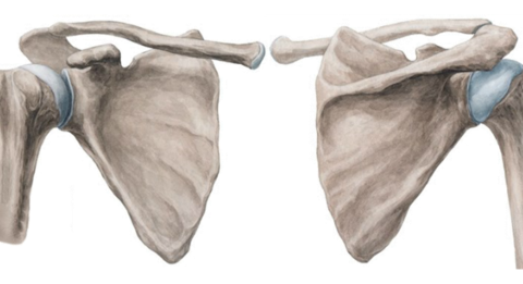
Scapula
By : zuhair Mohamed ammarType
It is flat bone and it has a triangular shape, roughly surface.
It forms the shoulder girdle.
It is placed on the posterolateral aspect of the thoracic cage between second rib and seventh rib.
•the scapula has two articulated sites with two bones:
1-with clavicle at the acromion process
2-with humerus bone at the glenoid process
It forms the shoulder girdle
Note
(with the clavicle at the sternoclavicular joint and the acromioclavicular joint )It is placed on the posterolateral aspect of the thoracic cage between second rib and seventh rib.
•the scapula has two articulated sites with two bones:
1-with clavicle at the acromion process
2-with humerus bone at the glenoid process
Angles ,borders and surfaces
The scapula has:
A)Three angles
1.Superior angle
2.Inferior angle
3.Lateral angle . See figure 1
B)Two surfaces
1.Anterior
2.Posterior
C)Three borders
1.Medial (vertebrae)
2.Lateral (axilla)
3. Superior
Note: Determination of the superior border is done by the suprascapular notch.
A)Three angles
1.Superior angle
2.Inferior angle
3.Lateral angle . See figure 1
B)Two surfaces
1.Anterior
2.Posterior
C)Three borders
1.Medial (vertebrae)
2.Lateral (axilla)
3. Superior
Note
( this border is divided by suprascapular notch into two thirds: 1-Medial two thirds 2-Lateral third)Note: Determination of the superior border is done by the suprascapular notch.
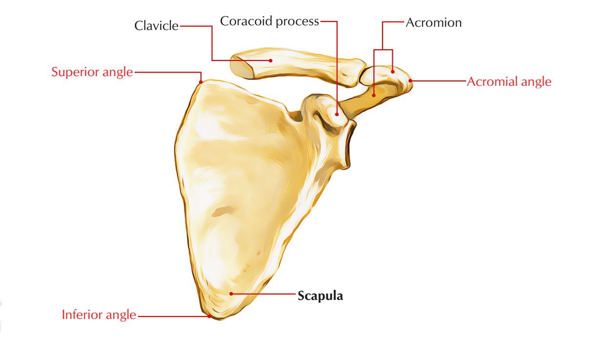

The posterior surface of the scapula
At the posterior surface there is a spine. And this spine:
1- can be used as a mark to determine the position of the scapula.
2- can be used as the medial point of attachment of the deltoid.
The spine and acromion serve as levers for the attached muscles, particularly the trapezius.
This spine divides the posterior surface into two parts:
1-Infraspinous fossa(below the spine)
2-Supraspinous fossa (above to spine)
• And this spine ends to form the acromial process
1- can be used as a mark to determine the position of the scapula.
2- can be used as the medial point of attachment of the deltoid.
The spine and acromion serve as levers for the attached muscles, particularly the trapezius.
This spine divides the posterior surface into two parts:
1-Infraspinous fossa(below the spine)
2-Supraspinous fossa (above to spine)
• And this spine ends to form the acromial process
Note
(the articulation part for clavicle and have attachments for some muscles)The Anterior surface of the scapula
At the anterior surface there is :
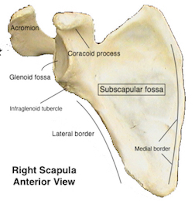
1.The subscapular fossa ( named according to the subscapularis muscles)
2.The prominent suprascapular notch . see figure 2
It is located at the lateral end of superior border.
Forms a pathway for important structures (suprascapular nerve and artery together accompanied ).
It is closed by a ligament.
It is transformed into foramen by the ligament.
It can be damaged and so affect the nerve below(suprascapular nerve)
1.The subscapular fossa ( named according to the subscapularis muscles)
2.The prominent suprascapular notch . see figure 2
It is located at the lateral end of superior border.
Forms a pathway for important structures (suprascapular nerve and artery together accompanied ).
It is closed by a ligament.
It is transformed into foramen by the ligament.
It can be damaged and so affect the nerve below(suprascapular nerve)
spinoglenoid notch

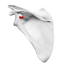
See figure 3.
This notch is placed at the lateral border of the spine and glenoid tubercle.
It is transformed into foramen by the ligament and this foramen acts as a pathway for suprascapular vessels from supraspinous fossa to infraspinous fossa .
This notch is placed at the lateral border of the spine and glenoid tubercle.
It is transformed into foramen by the ligament and this foramen acts as a pathway for suprascapular vessels from supraspinous fossa to infraspinous fossa .
Acromion process
This process forms the articulation surface for clavicle to form the acromioclavicular joint.
It is projected forward and slightly upward .
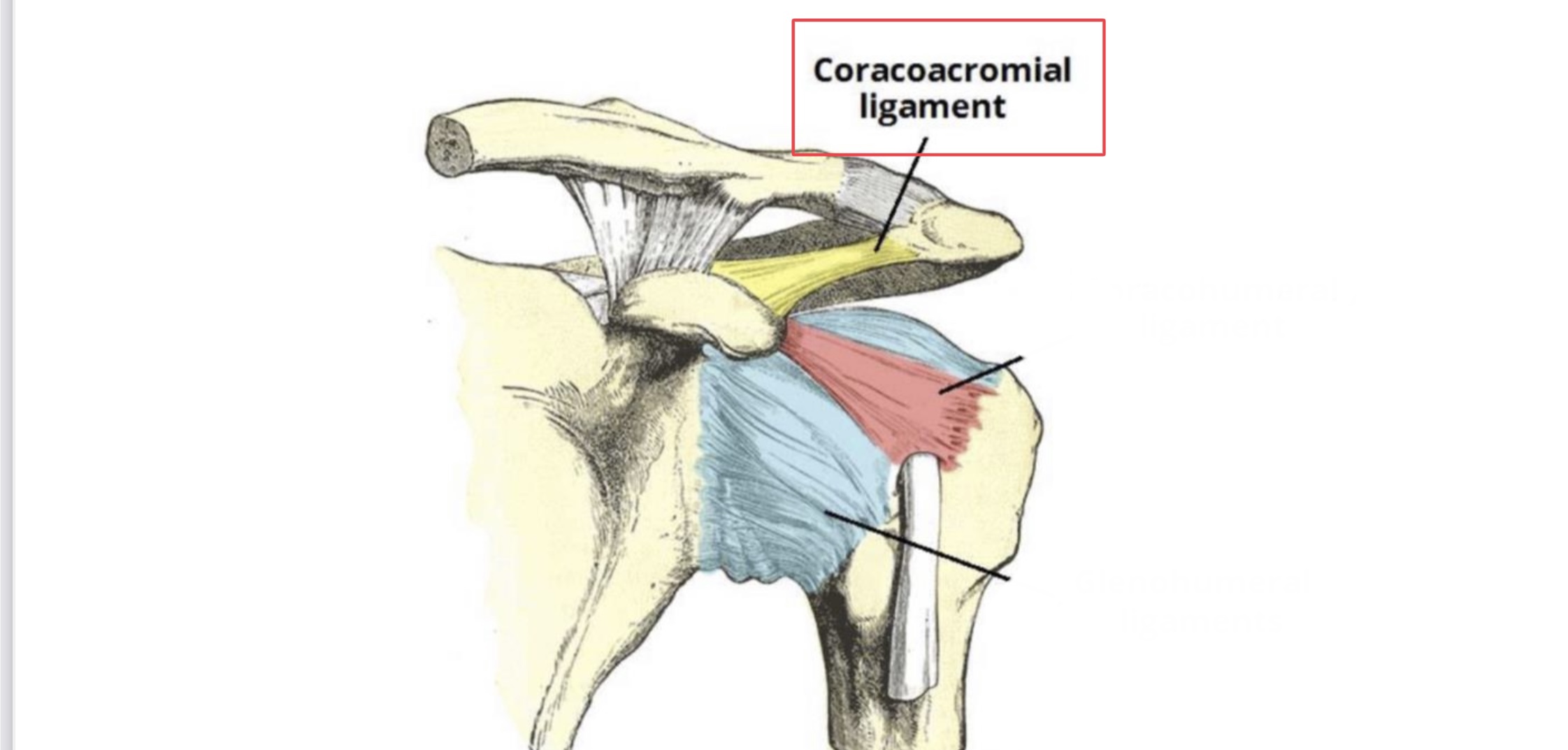
Has some important ligaments :
1-Superior & inferior acromioclavicular ligaments
2-Accessory ligament for the shoulder joint is (coracoacromial ligament)
See figure 4
Note
(this joint represents a part of the shoulder girdle)It is projected forward and slightly upward .
Has some important ligaments :
1-Superior & inferior acromioclavicular ligaments
2-Accessory ligament for the shoulder joint is (coracoacromial ligament)
See figure 4
Coracoid process
This process is projected forward and slightly laterally .
Has upper and lower surfaces.
Has medial and lateral borders.
Has an attachment point for some muscles and ligaments as:
Has upper and lower surfaces.
Has medial and lateral borders.
Has an attachment point for some muscles and ligaments as:
A)Ligaments
1-Coracoclavicular ligament(between clavicle and coracoid process)
2-Coracohumeral ligament (between coracoid process & greater tuberosity)
3-Coracoacromial ligament
4-Conoid ligament (between clavicle and coracoid process )
5-Trapezoid ligament (between clavicle and coracoid process)
1-Coracoclavicular ligament(between clavicle and coracoid process)
2-Coracohumeral ligament (between coracoid process & greater tuberosity)
3-Coracoacromial ligament
4-Conoid ligament (between clavicle and coracoid process )
5-Trapezoid ligament (between clavicle and coracoid process)
B)Muscles
1-Biceps m.
2-Coracobrachialis m.
3-Pectoralis minor m.
4- Subscapularis m.
1-Biceps m.
2-Coracobrachialis m.
3-Pectoralis minor m.
4- Subscapularis m.
Note
(It passes under the coracoid process )Notes
Neurovascular structures enter the upper limb medial to the coracoid process.
During treatment of coracoid related injuries, the musculocutaneous nerve and its branches should be identified and protected.
The surgeon should stay lateral to the coracoid instead of medial in order to avoid these neurovascular structures injuries
See figure 5
During treatment of coracoid related injuries, the musculocutaneous nerve and its branches should be identified and protected.
The surgeon should stay lateral to the coracoid instead of medial in order to avoid these neurovascular structures injuries
See figure 5

Supraspinous fossa

See figure 6
This is the superior part of the spine at the posterior surface of scapula.
It is separated from the infraspinous fossa by the spine of scapula .
It has a concave surface.
Superomeidally, there is a thicker area called the lateral angle.
At the lateral end, there is a glenoid cavity .
This fossa is named according to the presence of the supraspinatus muscle near of it.
There is a nerve(suprascapular nerve)enters the supraspinous fossa from the suprascapular notch then descends to infraspinous fossa. see figure 7
This is the superior part of the spine at the posterior surface of scapula.
It is separated from the infraspinous fossa by the spine of scapula .
It has a concave surface.
Superomeidally, there is a thicker area called the lateral angle.
At the lateral end, there is a glenoid cavity .
This fossa is named according to the presence of the supraspinatus muscle near of it.
There is a nerve(suprascapular nerve)enters the supraspinous fossa from the suprascapular notch then descends to infraspinous fossa. see figure 7

Infraspinous fossa
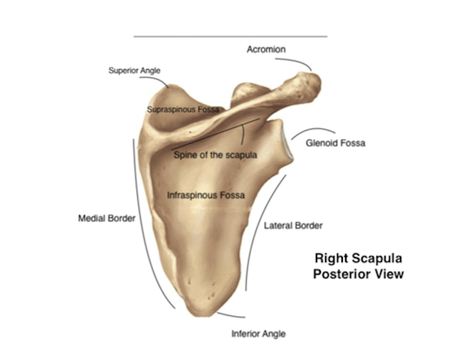
It is located at the posterior surface of scapula under the spine.
It has a triangular shape.
It is a site of some important muscle .
It has a notch spinoglenoid notch that connects the two fossae(supraspinous fossa and infraspinous fossa)
It is a very important area (functional point ) because it has the origin of three muscles (3 muscles of rotator cuff muscles):
1-Infraspinatus m.
2-Teres minor m.
3-Teres major m.
It has a triangular shape.
It is a site of some important muscle .
It has a notch spinoglenoid notch that connects the two fossae(supraspinous fossa and infraspinous fossa)
It is a very important area (functional point ) because it has the origin of three muscles (3 muscles of rotator cuff muscles):
1-Infraspinatus m.
2-Teres minor m.
3-Teres major m.
Subscapular fossa
This is the largest area of scapula at the anterior surface of scapula . See figure 9
It has three angles and three borders.
It is also a functional point because it has origin of the remaining muscle of the rotator cuff muscles which the subscapularis muscle. see figure 10
It has three angles and three borders.
It is also a functional point because it has origin of the remaining muscle of the rotator cuff muscles which the subscapularis muscle. see figure 10

Glenoid cavity

It is located at the lateral aspect of scapula (at lateral angle).
It makes a part of important joint called a shoulder joint. See figure 11
The meaning of glenoid in Greek is socket and it means the receiver area (receives the head of humerus to form a shoulder joint).
There are two tubercles around the glenoid cavity :
1.Supraglenoid tubercle
2.Infraglenoid tubercle
In these two tubercles, there are two important tendons pass:
1.Tendon of long head of biceps m. on supraglenoid tubercle
2.Long head of triceps m. on infraglenoid tubercle
It makes a part of important joint called a shoulder joint. See figure 11
The meaning of glenoid in Greek is socket and it means the receiver area (receives the head of humerus to form a shoulder joint).
There are two tubercles around the glenoid cavity :
1.Supraglenoid tubercle
2.Infraglenoid tubercle
In these two tubercles, there are two important tendons pass:
1.Tendon of long head of biceps m. on supraglenoid tubercle
2.Long head of triceps m. on infraglenoid tubercle
Clinical notes
The scapula needs a very strong damage to get fractured like car accident because the scapula is surrounded by muscles from each side.
The scapula can be changed or get problems due to:
1.position
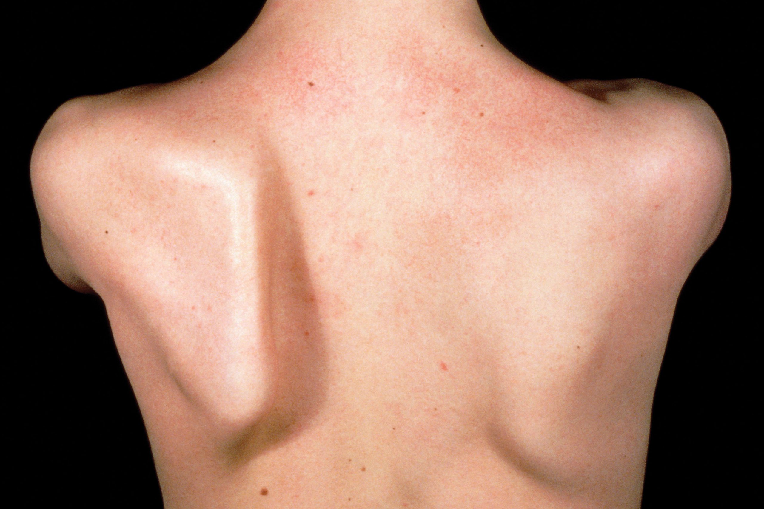
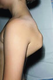
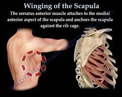
2.Muscles that surround the scapula like trapezius and serratus anterior muscles. When these muscles are paralyzed, the scapula will change its position (winged scapula) because the main action of these muscles is retraction and when they get paralyzed they quit their main action then the scapula will be protracted and so named a winged scapula .
The scapula can be changed or get problems due to:
1.position
2.Muscles that surround the scapula like trapezius and serratus anterior muscles. When these muscles are paralyzed, the scapula will change its position (winged scapula) because the main action of these muscles is retraction and when they get paralyzed they quit their main action then the scapula will be protracted and so named a winged scapula .
Distinguishing the side of scapula
To determination the side of scapula some steps should be done:
1. Imagining that the glenoid cavity will be articulated with humerus (it means it should be putted laterally )
2. Then putting the spine at posterior surface
1. Imagining that the glenoid cavity will be articulated with humerus (it means it should be putted laterally )
2. Then putting the spine at posterior surface
References
1. Dr. Lawrence E. Wineski,PhD;SNELLIS CLINICAL ANATOMY BY REGIONS 10th edition;ch3,p.89
2. Keith L. Moore , Arthur F. Dalley A. M. R. Agur; Moore clinically oriented anatomy 7th edition;ch.6,p.675
3. Richard Drake ,PhD , A. Wayne Vogl ,PhD ,Adam W. M. Mitchell ,MB BS FRCS FRCR , Richard Tibbitts , Paul Richardson; Gray's Atlas of Anatomy (Gray's Anatomy) 2nd Edition;p. 690
4. Sameh Doss, PhD; Bones of the Shoulder or Pectoral Girdle; ANATOMY HAND-OUT /UPPER LIM;p.5
5. The Structure Of The Scapula; interactive biology ;www.interactive-biology.com/4216/075-the-structure-of- the-scapula/
6. Basic Shoulder Anatomy; Cedars-Sinai Kerlan-Jobe Institute; https://tinyurl.com/y74ads7k
2. Keith L. Moore , Arthur F. Dalley A. M. R. Agur; Moore clinically oriented anatomy 7th edition;ch.6,p.675
3. Richard Drake ,PhD , A. Wayne Vogl ,PhD ,Adam W. M. Mitchell ,MB BS FRCS FRCR , Richard Tibbitts , Paul Richardson; Gray's Atlas of Anatomy (Gray's Anatomy) 2nd Edition;p. 690
4. Sameh Doss, PhD; Bones of the Shoulder or Pectoral Girdle; ANATOMY HAND-OUT /UPPER LIM;p.5
5. The Structure Of The Scapula; interactive biology ;www.interactive-biology.com/4216/075-the-structure-of- the-scapula/
6. Basic Shoulder Anatomy; Cedars-Sinai Kerlan-Jobe Institute; https://tinyurl.com/y74ads7k