
Histology of Cardiovascular system
By : Omar Al-abdrabaElastic arteries
Like : aorta.
It’s composed of:
1- tunica intima : it’s the inner layer and composed of endothelium, subendothelial C.T and internal elastic membrane (internal elastic lamina) which appears as dark purple wave between tunica intima and tunica media in fig1.
2- tunica media : it’s the middle layer and is the thickest one between the 3 layers in elastic arteries(fig2).
Its composed of alternating layers of sheets of elastic tissue (appears dark purple in fig3) and smooth muscles (appears unstained in fig3 )arranged circumferentially.
3- tunica adventitia : it’s the outer layer composed of dense irregular connective tissue.
It’s usually less than the thickness of tunica media in elastic arteries(fig2).
*Vasa vasorum : blood vessels that supply the tunica adventitia and tunica media (fig4).
Used slides from histology guide:
1-MHS 244
2-MH 065-066
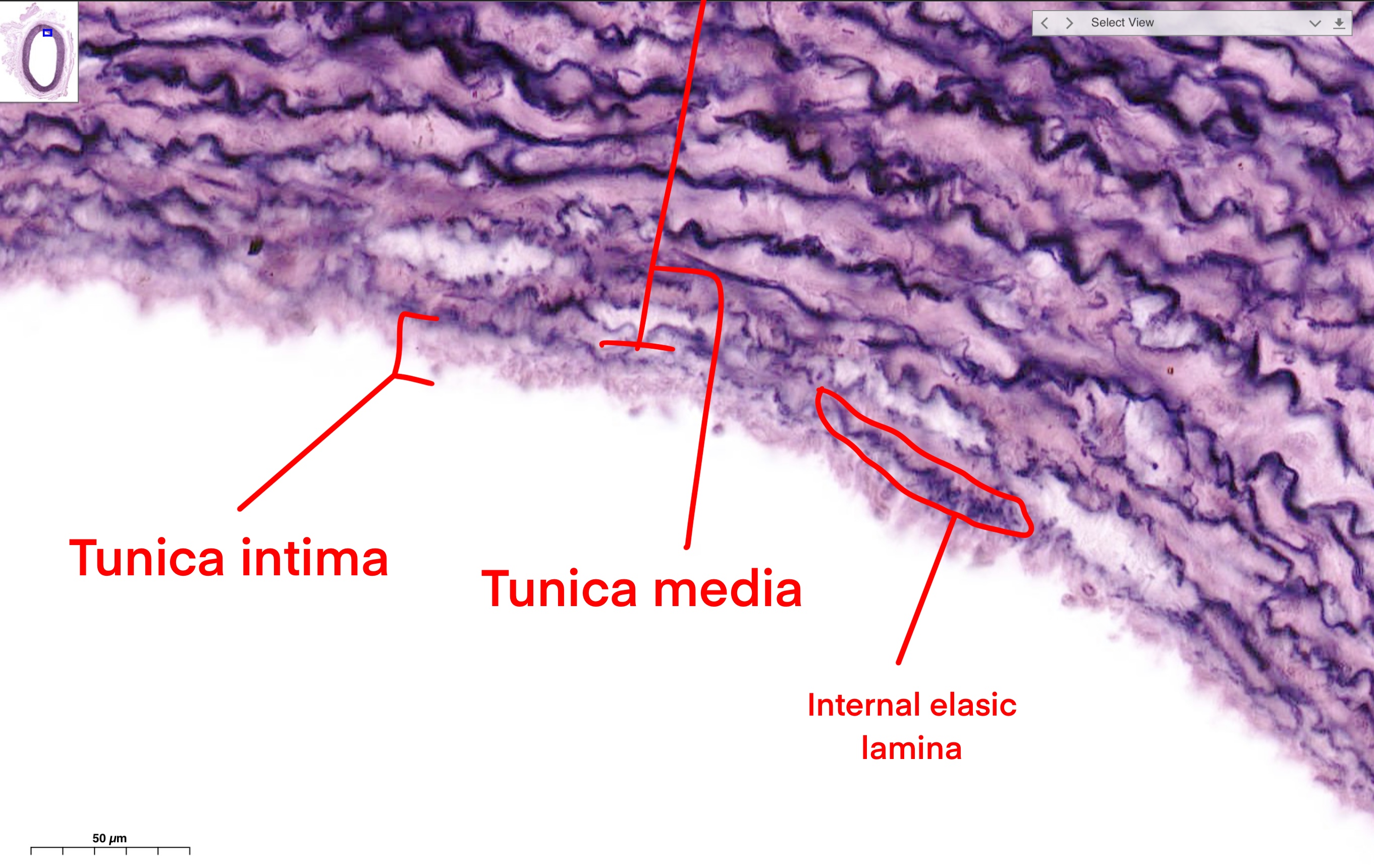

From slide ( MHS244 )
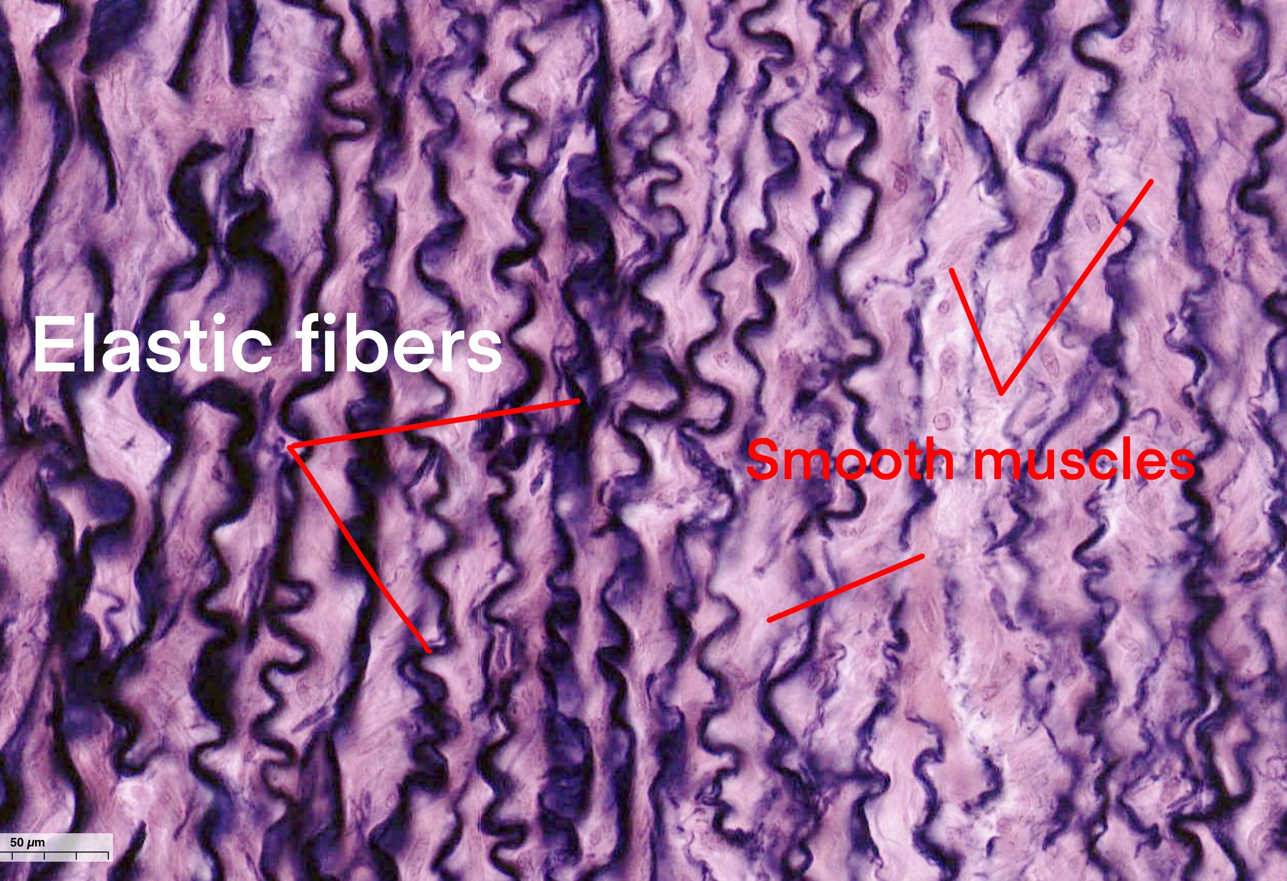
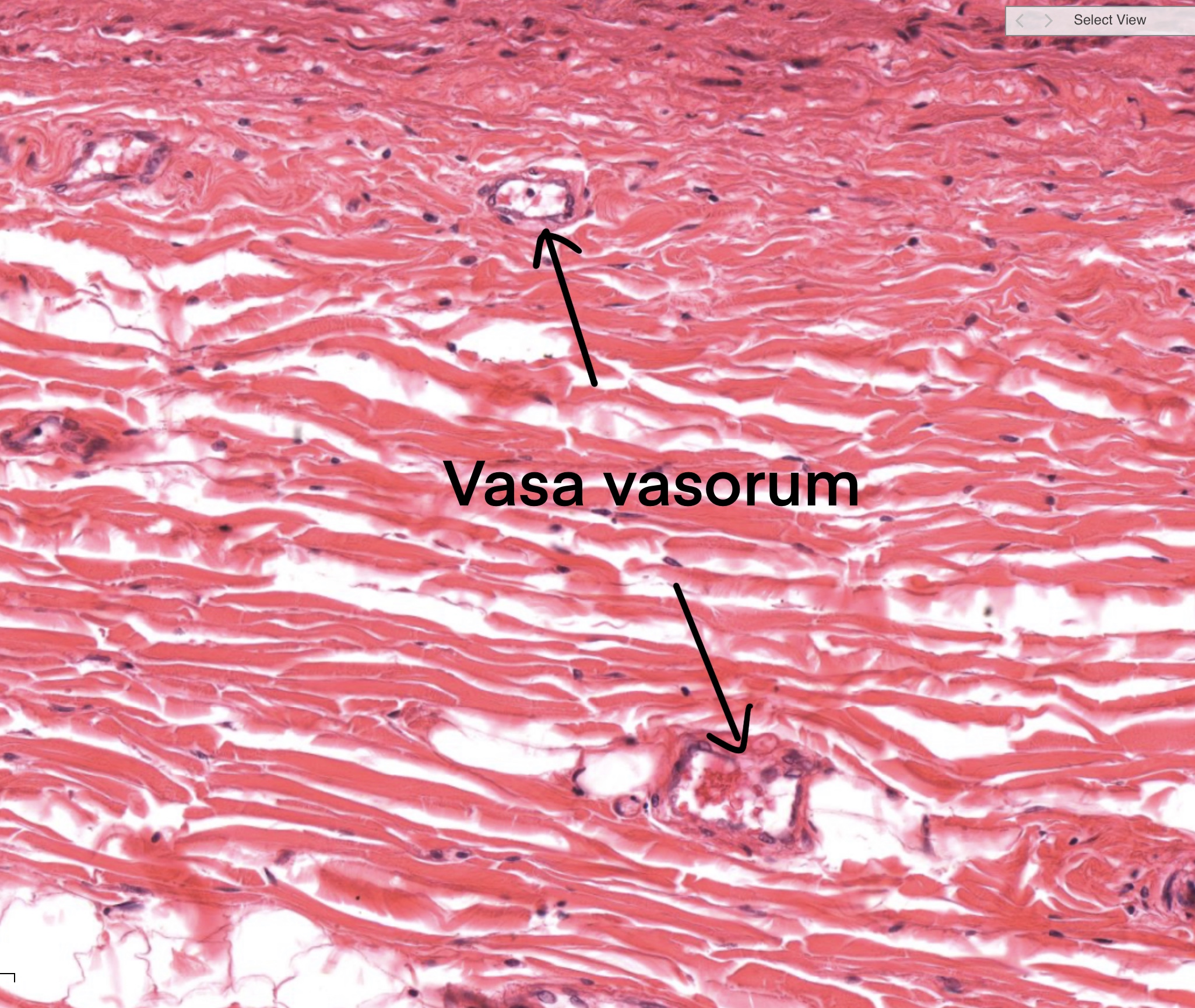
From slide ( MHS 244 )
Large veins
Like:vena cava, it’s wall is thinner than the wall of aorta.
1-tunica intima : it’s the inner layer composed of endothelium + thin layer of subendothelial C.T..(fig5).
2-tunica media : it’s the middle layer consists of 3-8 smooth muscle layers (fig5).
3- Tunica Adventitia : it’s the outer layer composed of irregular dense connective tissue D.C.T containing longitudinal arrangements of smooth muscle + collagen fibers (fig5 and fig6).
*Vasa Vasorum : blood vessels that supply the tunica adventitia and tunica media(fig7).
Used slides from histology guide:
1-MH 065-066
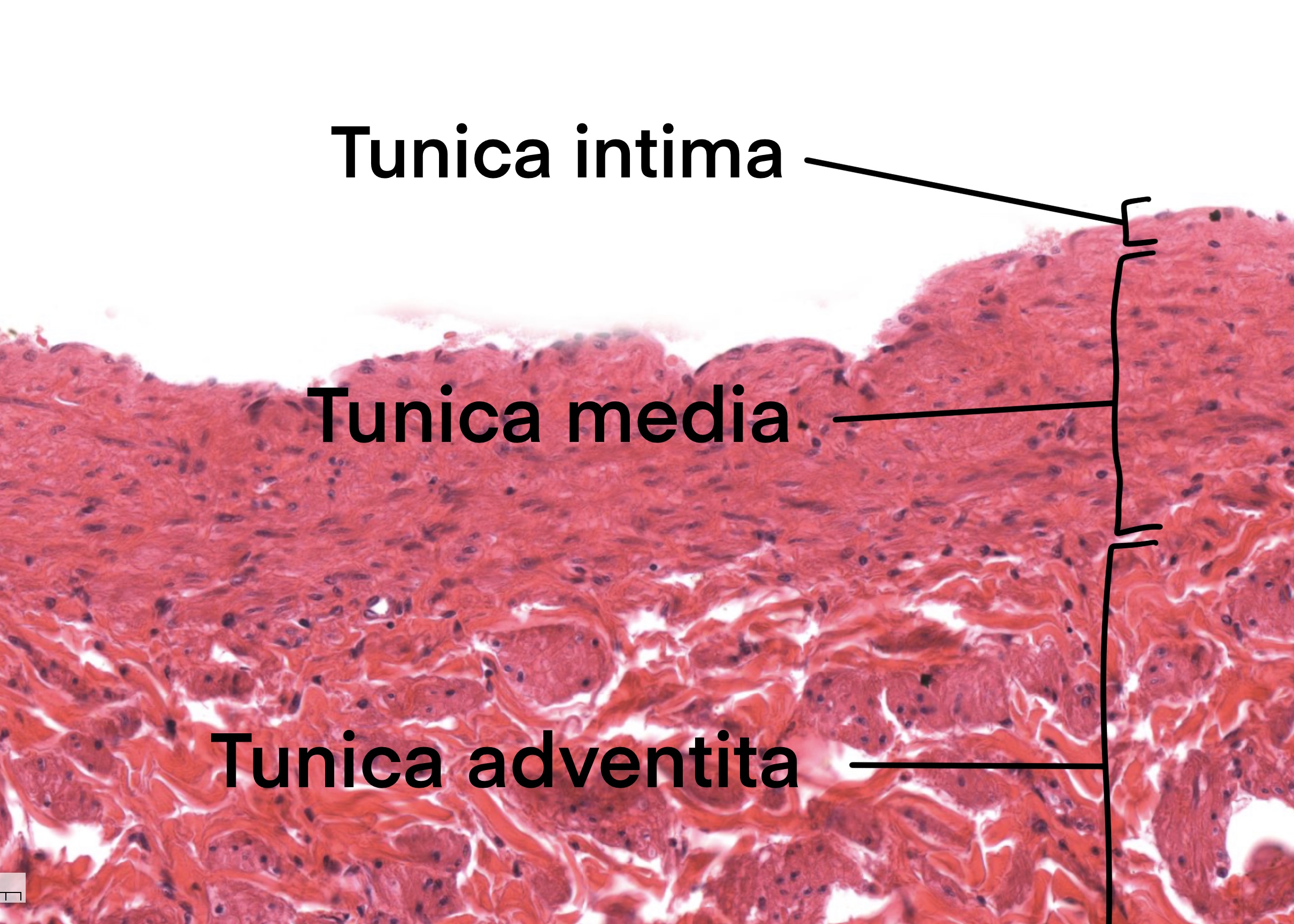

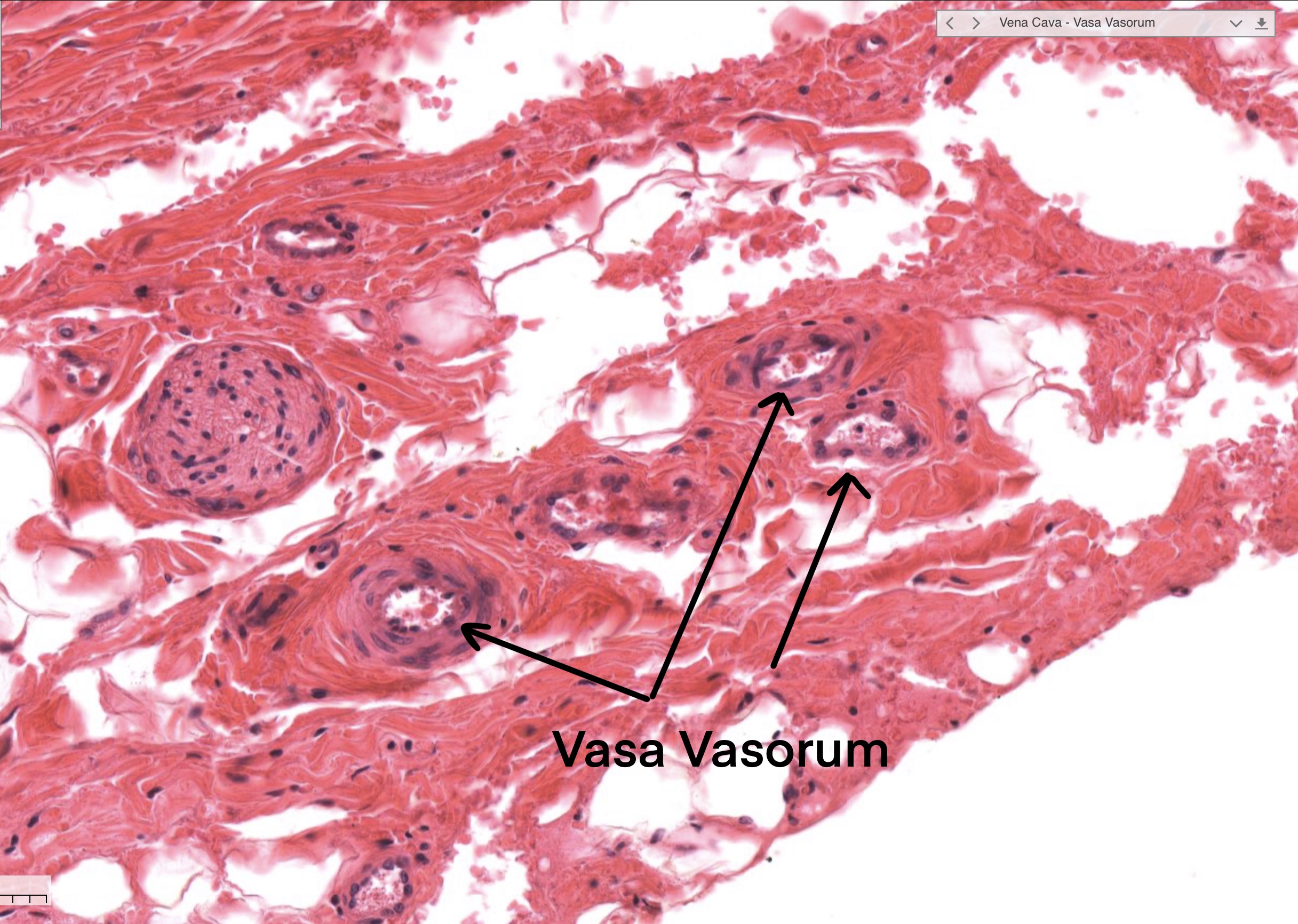
Slide (MH 065-066 )
Muscular arteries and medium veins
Muscular Artery has a tunica intima with a prominent internal elastic lamina and tunica media with a prominent smooth muscle component.
1-Tunica Intima: inner layer composed of the endothelium, subendothelial C.T, and a prominent internal elastic lamina.
The internal elastic lamina appears as a wavy band.
2-Tunica Media: middle layer composed mostly of circumferentially arranged S.M smooth muscle.
3-Tunica Adventitia: outer layer composed of well-organized dense irregular C.T connective tissue.
Used slides from histology guide:
1-MH 061-062
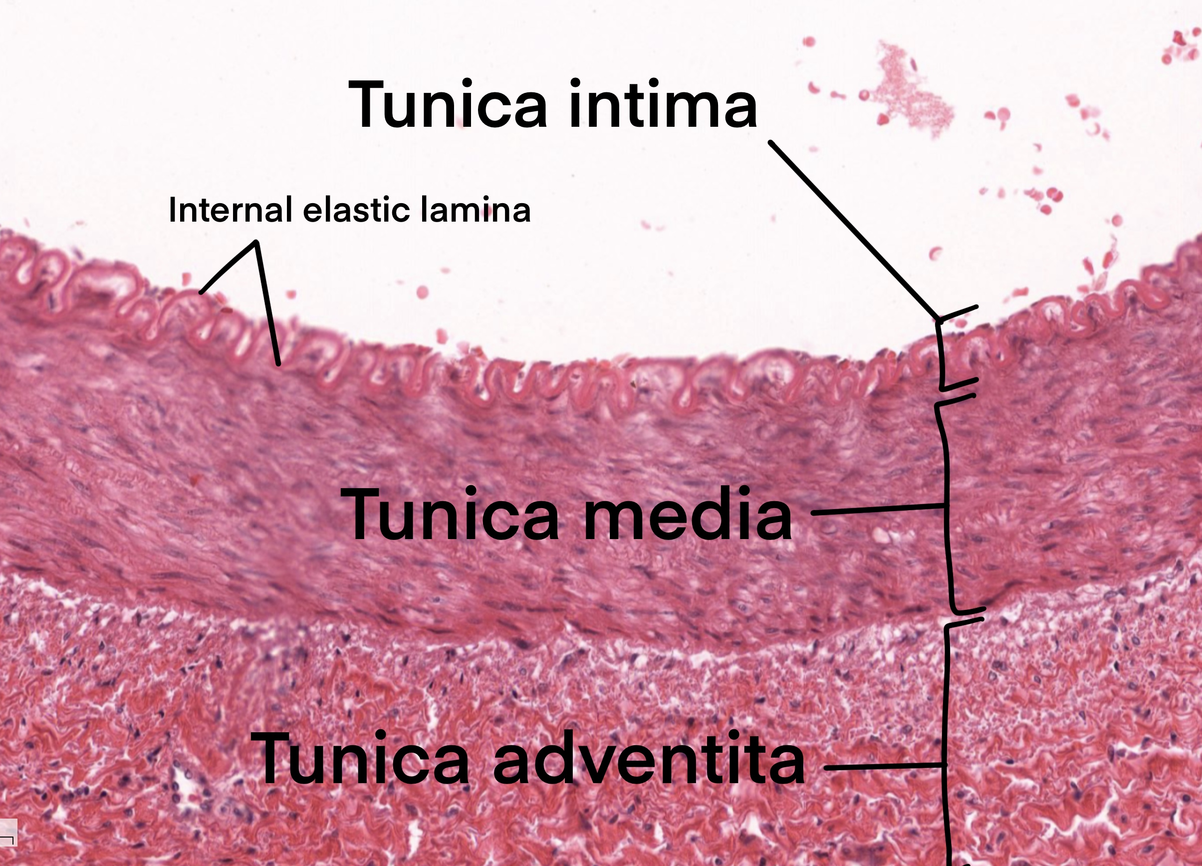
Medium Vein has less distinct layers than the artery.
1-Tunica Intima: inner layer composed of the endothelium, subendothelial CT.
2-Tunica Media: middle layer of only a few smooth muscle layers circumferentially arranged.
3-Tunica Adventitia: outer layer composed of (D.C.T+ SM) dense irregular connective tissue & scattered smooth muscle.
Used slides from histology guide:
1-MH 061-062
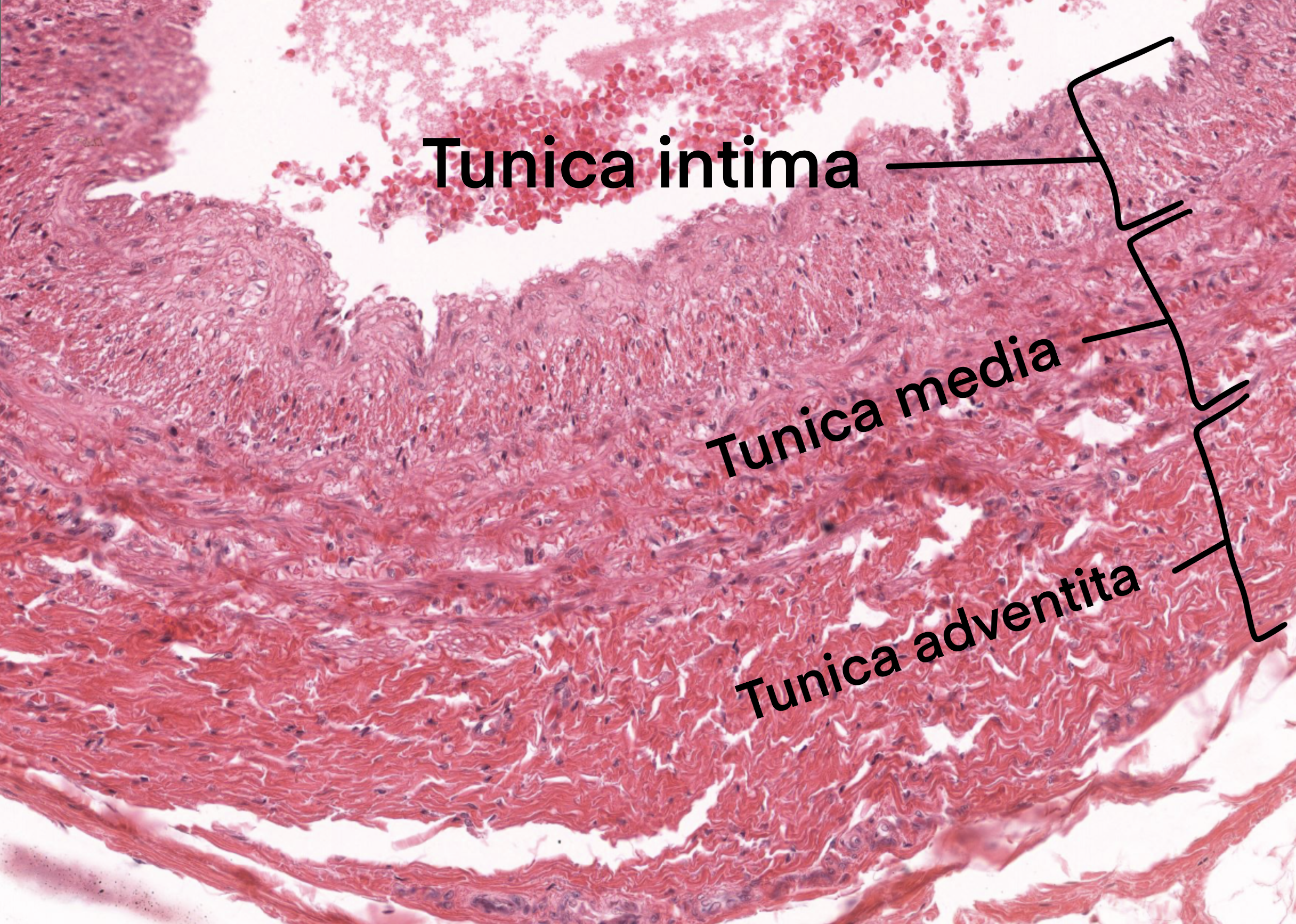
Slide ( MH 061-062 )
Arterioles and venules
Arterioles is small diameter blood vessels.
Their wall consists of endothelial cells, one or two layers of smooth muscle, and a thin layer of collagen fibers.
The inner elastic lamina is usually absent from smaller arterioles (you will se the inner elastic lamina in the areriol on the right while missing on the smaller arteriol on the left in fig8 ).
Used slides from histology guide:
1-MH 024-025


Slide ( MH 024-025 )
Venules are small diameter blood vessel that allows blood to return from capillary beds to veins.
Their wall is composed of an endothelial cell, one or two layers of smooth muscle, and very thin adventitia.
Used slides from histology guide:
1-MH 024-025
Their wall is composed of an endothelial cell, one or two layers of smooth muscle, and very thin adventitia.
Used slides from histology guide:
1-MH 024-025
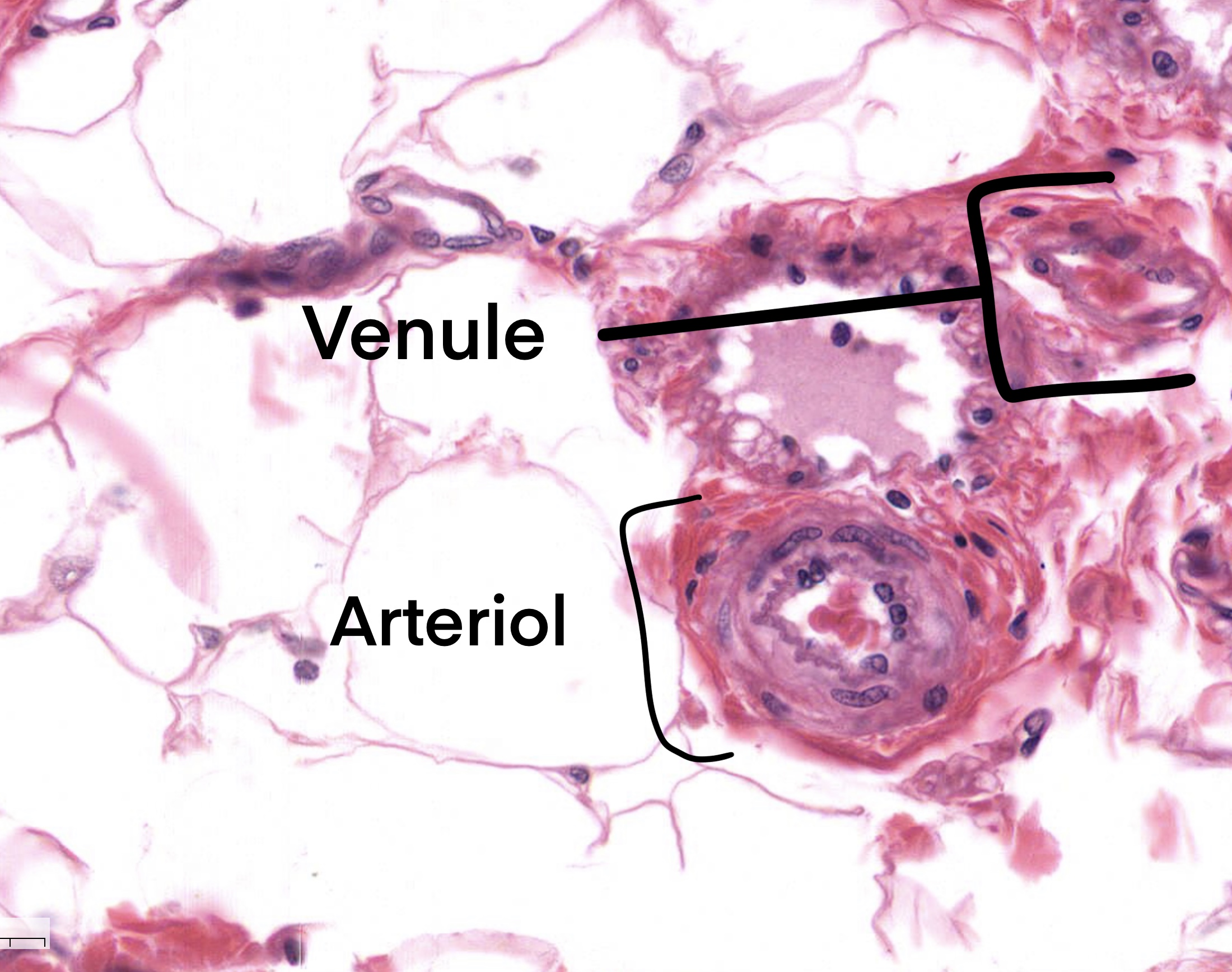
Slide ( MH 024-025 )
Capillaries and Sinusoids
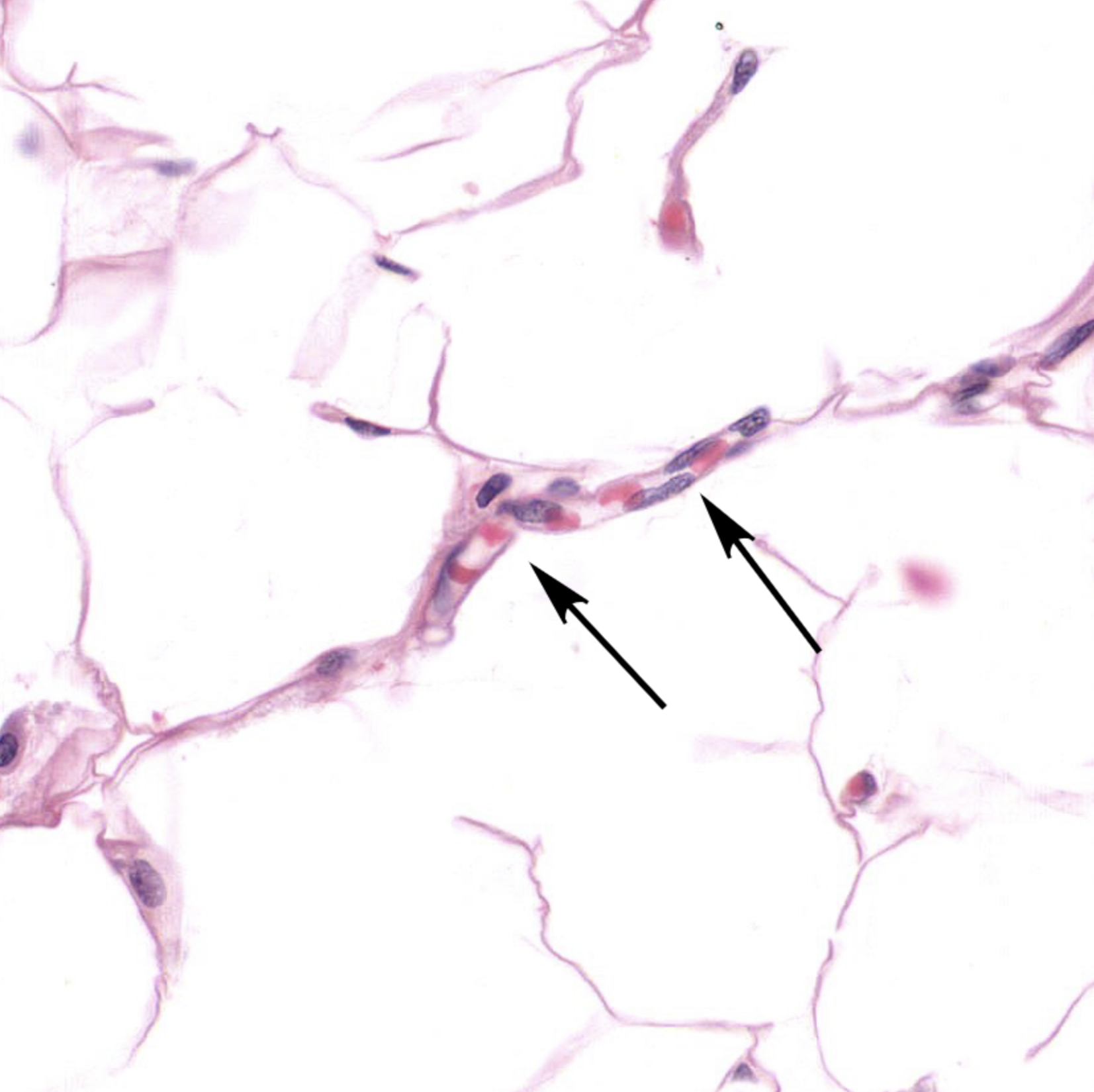
Capillaries are the smallest diameter blood vessels.
Their wall is a one-layer endothelium.
A red blood cell in the lumen next to the nucleus of an endothelial cell shown in fig9
Longitudinal section of a capillary containing red blood cells. The flattened nuclei of endothelial cells are visible in fig10.
Used slides from histology guide:
1-MH024
Their wall is a one-layer endothelium.
A red blood cell in the lumen next to the nucleus of an endothelial cell shown in fig9
Longitudinal section of a capillary containing red blood cells. The flattened nuclei of endothelial cells are visible in fig10.
Used slides from histology guide:
1-MH024

Slide ( MH24 )
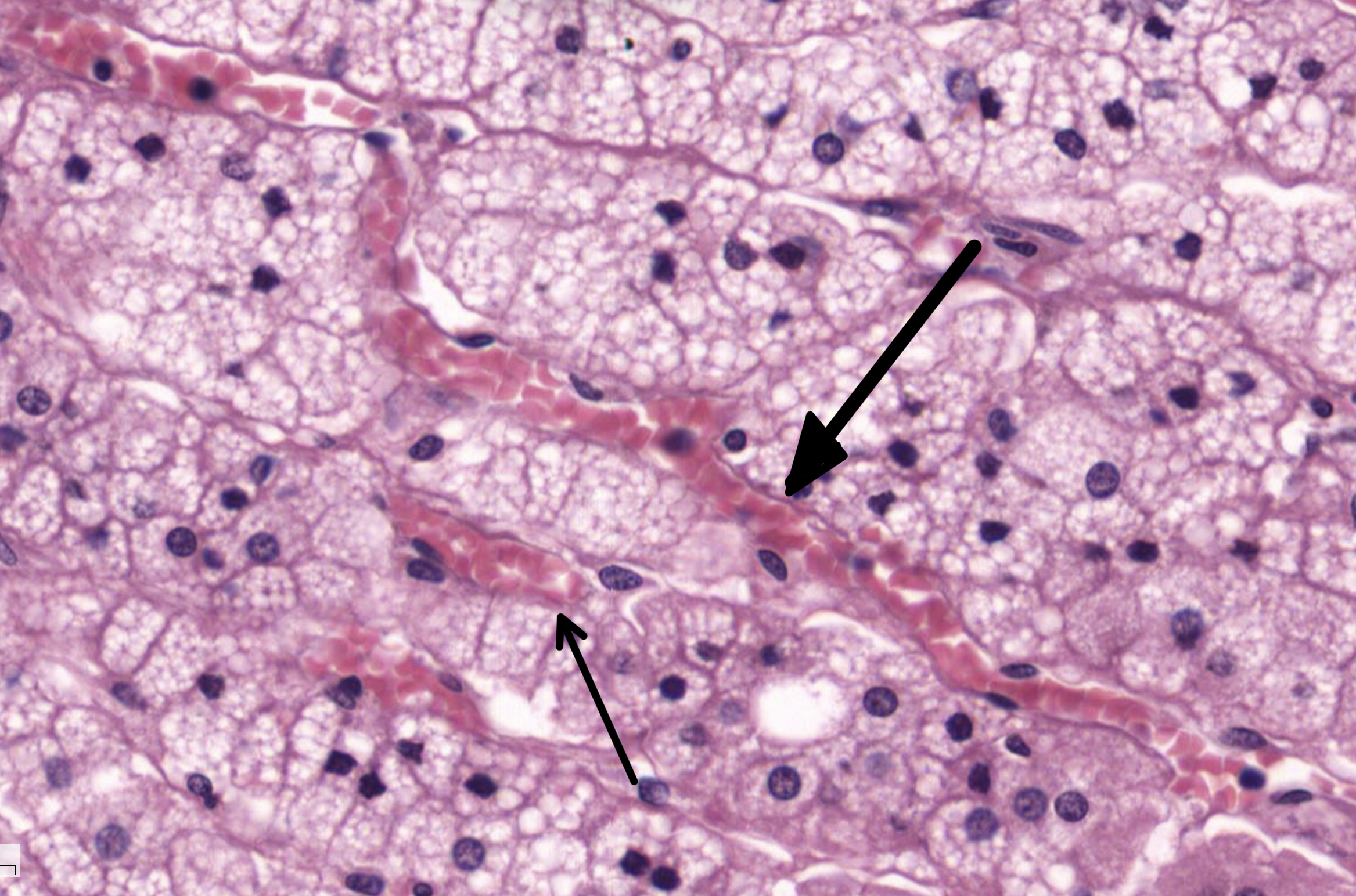
Sinusoidal capillaries (or sinusoids) have a discontinuous endothelium and basement membrane.
Sinusoidal capillaries are found in a number of organs including the liver, spleen, and bone marrow Sinusoids are also found in the adrenal gland.
The adrenal cortex is richly supplied by a network of sinusoids.
Used slides from histology guide:
1-MH115a
Sinusoidal capillaries are found in a number of organs including the liver, spleen, and bone marrow Sinusoids are also found in the adrenal gland.
The adrenal cortex is richly supplied by a network of sinusoids.
Used slides from histology guide:
1-MH115a
Slide ( MH 115a )
