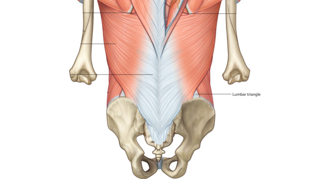
Lumbar triangle
By : Amna Mohammed
The human body has two lumbar triangles:
1. Superior lumbar triangle
It is also known as Grynfeltt-Lesshaft .
Location
Posterolateral aspect of the abdomen region.
Shape
It has a triangle shape.
Boundaries
It has three borders:
Superiorly: Lower border of 12th rib
Medially: Quadratus oblique m.
Laterally: Posterior border of Internal oblique m.
The floor: Transversalis fascia
The roof: External abdominal oblique m.
Superiorly: Lower border of 12th rib
Medially: Quadratus oblique m.
Laterally: Posterior border of Internal oblique m.
The floor: Transversalis fascia
The roof: External abdominal oblique m.
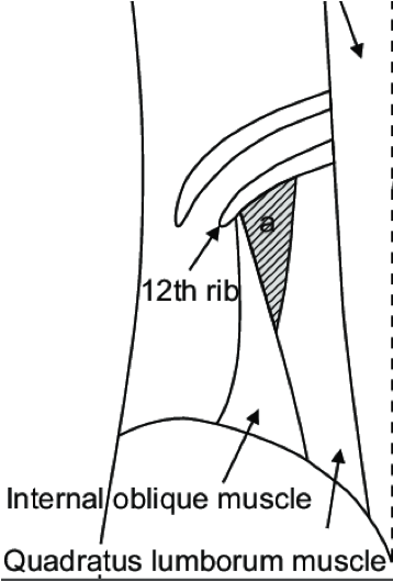
Posterior view
2. Inferior lumbar triangle
It is also known as Petit's triangle .
Location
Posterolateral aspect of the abdomen region.
Shape
It has a triangle shape.
Boundaries
It has three borders:
Inferiorly: Iliac crest
Medially(Posteriorly): Lateral border of latissimus dorsi m.
Laterally(Anteriorly): Posterior border of external abdominal oblique m.
The floor: Internal abdominal oblique m. & transversus abdominis m.
Inferiorly: Iliac crest
Medially(Posteriorly): Lateral border of latissimus dorsi m.
Laterally(Anteriorly): Posterior border of external abdominal oblique m.
The floor: Internal abdominal oblique m. & transversus abdominis m.
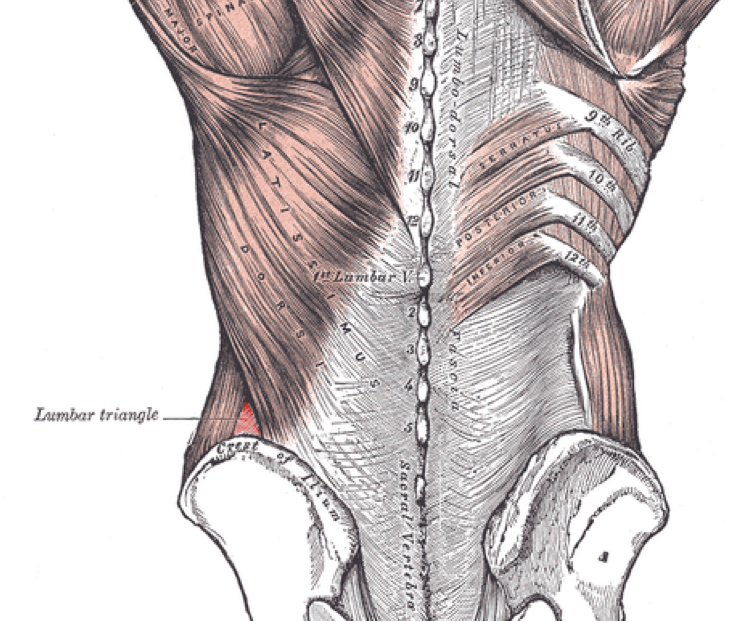
Posterior view
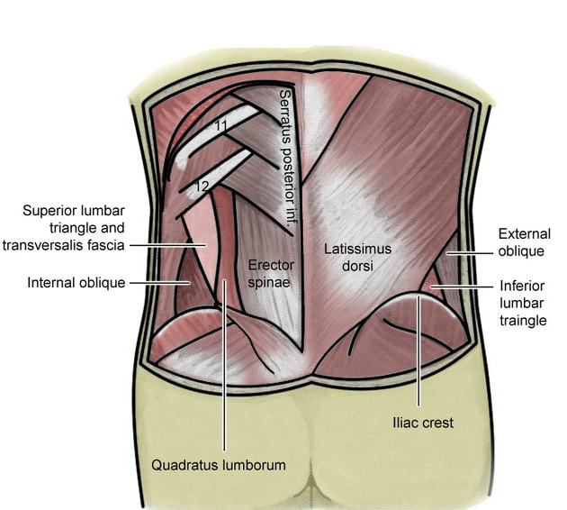
Posterior view
Clinical importance
Its clinical significance is that it is a weak site between the muscles, so it is prone to the occurrence of the lumbar hernia
Note
the protrusion of an organ or organ membrane through the wall of the cavity which contains it.Lumbar hernia
A condition in which structures protrude from the abdominal cavity posteriorly through the lumbar triangle.
It's common among:
1. 55-70 years old people.
2. Among men more than women.
Note: petit lumbar hernia is rarely more than Grynfeltt lumbar hernia.
These structures usually contain Intestinal fat (fat over the bowel), colon, small bowel, stomach, ovary, spleen, appendix, the greater omentum, urinary bladder, renal pelvis, gallbladder, liver, or kidney.
Note: Grynfeltt lumbar hernia is more respectable to trap a part of the intestine in some cases more than petit lumbar triangle because its neck size is small (narrow).
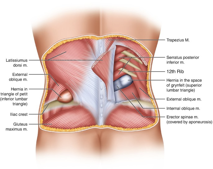
Posterior view, show the two type of Lumbar hernia.
The Possible causes of lumbar hernia
1. In childhood, a disorder in the musculoskeletal system.
2. In adolescence, There are two types of reasons:
1. Unprompted, without any causative agent like infection, trauma.
2. Prompted, there are causative agents such as cracking trauma, hepatic abscesses, Severe abdominal injuries, etc.
Note: some hazard agents that affect unprompted reasons such as age, rapid weight loss, muscular atrophy, chronic diseases etc
The Symptoms
1. Back pain which increases during coughing.
2. Lump on the side of the abdomen which disappears when lying down & It increases in size over time.
3. Abdomen spasms.
4. Debility in one leg or foot
2. Lump on the side of the abdomen which disappears when lying down & It increases in size over time.
3. Abdomen spasms.
4. Debility in one leg or foot
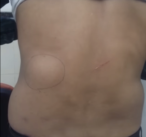
Lumbar hernia, show a lump on the lateral side of the abdomen, posterior view.
The Diagnosis
1. The specialist uses ultrasound to diagnose if the hernia contains intestine to hear the movement of the intestine through the hernia.
2. If the hernia contains an amount of fat, then the use of sound waves will be inefficient, so is resorted to the use of computerized tomography(it is good for distinguishing the hernia contents).
Note: sometimes may be misdiagnosed as lumbar back pain.
2. If the hernia contains an amount of fat, then the use of sound waves will be inefficient, so is resorted to the use of computerized tomography(it is good for distinguishing the hernia contents).
Note: sometimes may be misdiagnosed as lumbar back pain.
The Treatment
A lumbar hernia doesn’t need surgery always(if the symptoms are tenuous).
If the lumbar hernia is painful & acute . It can be treated surgically, as there are multiple techniques of surgery, the choice of which depends on the experience of the surgeon & the diameter of the hernia .
Here are some of the techniques used:
1. Repairing the hernia by retro-muscular or sublay prolene mesh as this can maintain the maximum overlap of healthy tissue with the implanted mesh material.
2. It can also be repaired using laparoscopic in case of uncomplicated lumbar hernias that can be performed with equivalent success.
3. Other techniques include anatomical closure, overlapping of the aponeuroses, use of musculofascial flaps, prosthetic meshes
The patient can be out after 4-6 days from the hospital after the procedure the surgery.
Note\ An appropriate surgical treatment should be planned to grant the patient:
1. Less time spent in the hospital
2. Decreased use of painkillers
3. Back to daily activities in a short time 4.Less chance of wound infection.
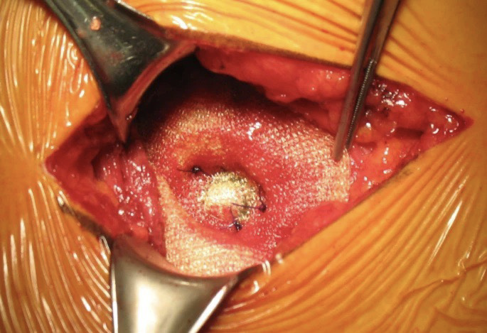
Prolene mesh , a way to treatment the lumbar hernia.
References
SNELL'S CLINICAL ANATOMY BY REGIONS 10th Edition
158,762
A primary idiopathic superior lumbar triangle hernia with congenital right scoliosis: A rare clinical presentation and management- PubMed- https://pubmed.ncbi.nlm.nih.gov/23776777/
Anatomical and surgical considerations on lumbar hernias- PubMed- https://pubmed.ncbi.nlm.nih.gov/19999919/
Lumbar Hernia- PubMed- https://www.ncbi.nlm.nih.gov/pmc/articles/PMC4921421/
Anatomical and surgical considerations on lumbar hernias- pubMed- https://pubmed.ncbi.nlm.nih.gov/19999919/
LUMBAR HERNIA TREATMENT- CORE SURGICAL- https://coresurgicalmd.com/lumbar-hernia/
Lumbar hernia- Radiopaedia- https://radiopaedia.org/articles/lumbar-hernia?lang=us
Lumbar hernia- mAnchester Surgical Clinic- https://www.manchestersurgicalclinic.com/conditions/hernia/lumbar-hernia
https://youtu.be/btG39rXs5CM
https://quizlet.com/222445179/anatomical-snuff-box-labeling-diagram/
https://astr.or.kr/ViewImage.php?Type=F&aid=612888&id=F4&afn=6037_ASTR_95_6_340&fn=_6037ASTR
https://radiopaedia.org/articles/superior-lumbar-triangle
https://en.wikipedia.org/wiki/Lumbar_triangle#/media/File:Lumbar_triangle.PNG
https://medizzy.com/feed/34261480
https://jmedicalcasereports.biomedcentral.com/articles/10.1186/1752-1947-3-9322/figures/4
