


AXILLARY REGION
By : عبدالله محمدShape:
(Armpit) of the axilla is pyramid-shaped area.
It is a space between the chest side and the upper part of the arm.
It is a space between the chest side and the upper part of the arm.
Function:
Functions of the axilla:
It is the only passageway of the blood,nerves,and lymph vessels from
the root of the neck to the upper limb.
It is the only passageway of the blood,nerves,and lymph vessels from
the root of the neck to the upper limb.
Boundaries of the axilla:
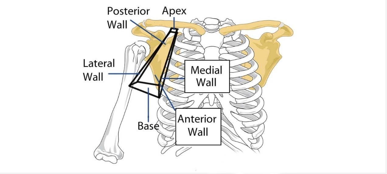
The axilla has 4 walls in addition to the apex and base.
The walls: (Anterior,posterior,medial,and lateral).
The walls: (Anterior,posterior,medial,and lateral).
Note
The shape and the size of the axilla is varieng and depends on the arm position.Base of axilla:
formed when the skin stretchs between the posterior and the
anterior walls of the axilla.
1-In front: the base of the axilla is formed by the anterior axillary fold
(formed by the pectoralis major muscle(the lower border))
2- Behind: the base of the axilla is formed by the posterior axillary fold (formed by the tendons of latissimus dorsi and teres major
muscles).
Apex of axilla:
it is narrow and triangular.
-It lies in the upper part of the axilla.
-It is directed medially and upward.
-It connects the posterior triangle of the neck to the axilla
(cervicoaxillary canal).
-It lies in the upper part of the axilla.
-It is directed medially and upward.
-It connects the posterior triangle of the neck to the axilla
(cervicoaxillary canal).
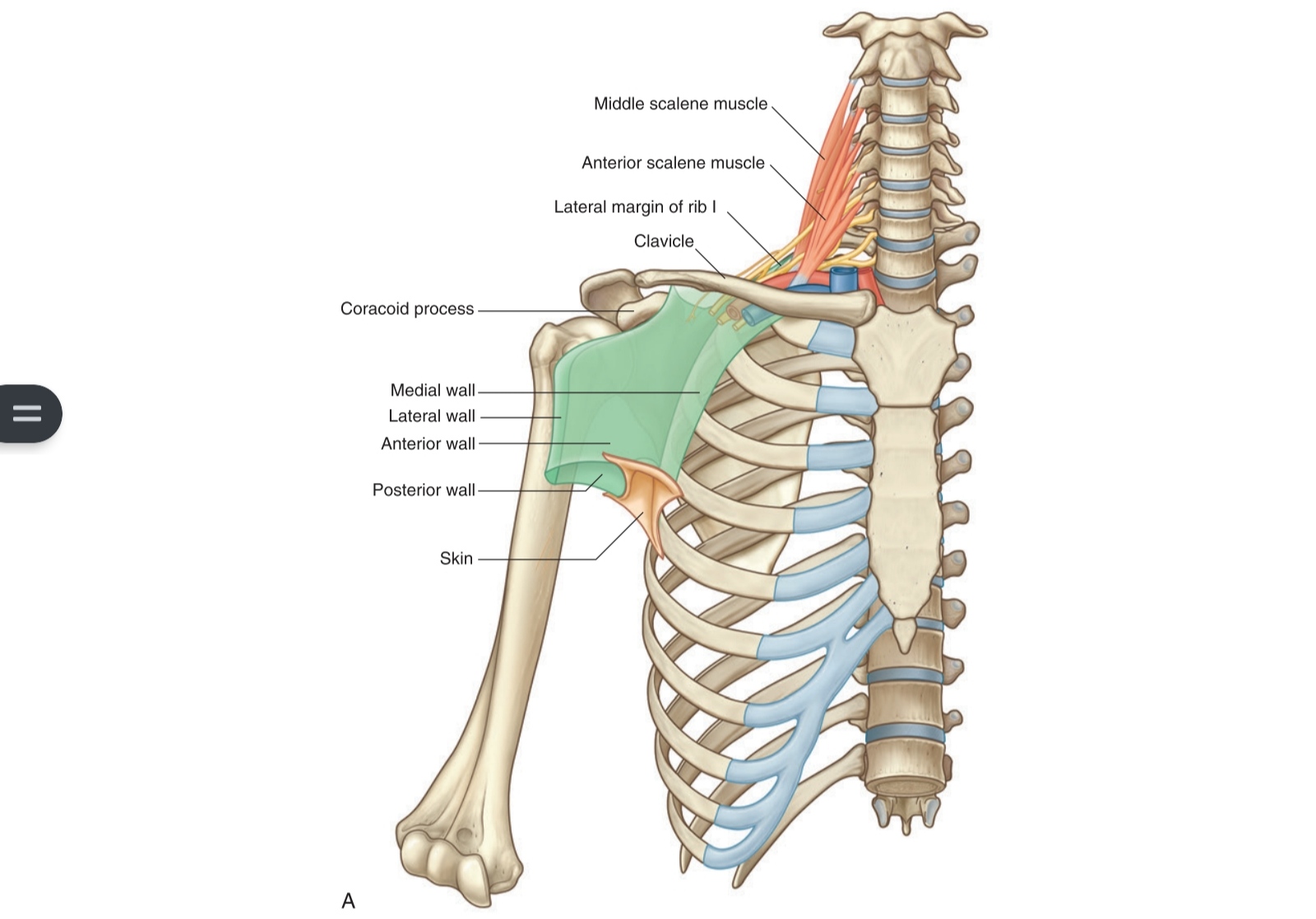
Boundaries of the apex:
1-Anteriorly:the middle third of the clavicle.
2-Posteriorly: the upper border of the scapula.
3-Medially:the outer border of the 1st rib
Note:the apex doesn't have a lateral boundary because it's a
trianglular-shaped .
2-Posteriorly: the upper border of the scapula.
3-Medially:the outer border of the 1st rib
Note:the apex doesn't have a lateral boundary because it's a
trianglular-shaped .
The walls of axilla:
1. Anterior wall is formed by:
A- pectoralis major muscle
B- pectoralis minor muscle
C-subclavius muscle
D-clavipectoral fascia
2. Posterior wall is formed by:
A-subscapularis muscle
B-latissimus dorsi muscle
C-teres major muscle
3. Medial wall is formed by:
A-serratus anterior muscle
B- the upper 4-5 ribs and the intercostal muscles
Note: The medial wall is wide .
4. Lateral wall is formed by:
A- coracobrachialis muscle
B-biceps brachii muscle
C-the bicipital groove of the humerus.
Note:the lateral wall is narrow .
Axillary sheath
It is the axillary fascia which encloses the axillary artery and the brachial plexus.
Note : the axillary sheath is a continuation of the prevertebral fascia
of the deep cervical fascia.
Note : the axillary vein is not one of the contents of the axillary sheath to
allow the vein to expand freely during the increased venous return.
Note : the axillary sheath is a continuation of the prevertebral fascia
of the deep cervical fascia.
Note : the axillary vein is not one of the contents of the axillary sheath to
allow the vein to expand freely during the increased venous return.

Passageways Exiting The Axilla
There are three main routes by which structures leave the axilla:
1- The main route of exit is immediately inferiorly and laterally, into the
upper limb. The contents of the axilla region leave by this method majority.
2- The quadrangular space:
It is a gap in the posterior wall of the axilla that's allowing access to
the posterior arm and shoulder region.
3-The clavipectoral triangle: It is an opening in the anterior wall of the
axilla,it is bounded by the
deltoid, clavicle and pectoralis major muscle. Via this triangle, the
cephalic vein enters the axilla, while the medial pectoral nerve and the
lateral pectoral nerve leave through it.
Axillary fascia
The axillary fascia is dense , especially:
1- below the deltoid muscle.
2- in the central third of the shoulder At the lateral margin of the
latissimus dorsi muscle.
it divides into:
two layers, which ensheaths the muscles, and are attached posteriorly to the
spinous processes of the thoracic vertebrae.
1- below the deltoid muscle.
2- in the central third of the shoulder At the lateral margin of the
latissimus dorsi muscle.
it divides into:
two layers, which ensheaths the muscles, and are attached posteriorly to the
spinous processes of the thoracic vertebrae.
Axillary folds
1- Anterior axillary fold:
A- is the lower border of anterior wall of axilla.
B- it is formed by the lower border of the pectoralis major muscle.
2- Posterior axillary fold:
A- is the lower border of posterior wall of axilla.
B-it is formed by both teres major and latissimus dorsi muscles.
A- is the lower border of anterior wall of axilla.
B- it is formed by the lower border of the pectoralis major muscle.
2- Posterior axillary fold:
A- is the lower border of posterior wall of axilla.
B-it is formed by both teres major and latissimus dorsi muscles.
Axillary lines
The axillary lines: are imaginary lines,
1- Anterior axillary line:it is the continuation of the anterior
axillary fold.
2- Posterior axillary line:it is the continuation of the posterior
axillary fold.
Note:the midaxillary line lies midway between the anterior and the
posterior axillary lines.
Note :nerve to serratus anterior muscle descends vertically in the
midaxillary line.
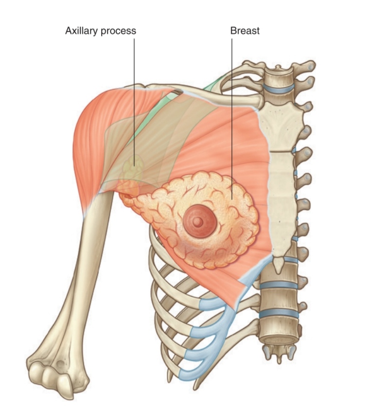
Axillary tail
an upward extension of the mammary gland into the axilla.
Note: only the females have the axillary tail, while males do not.
Note: only the females have the axillary tail, while males do not.
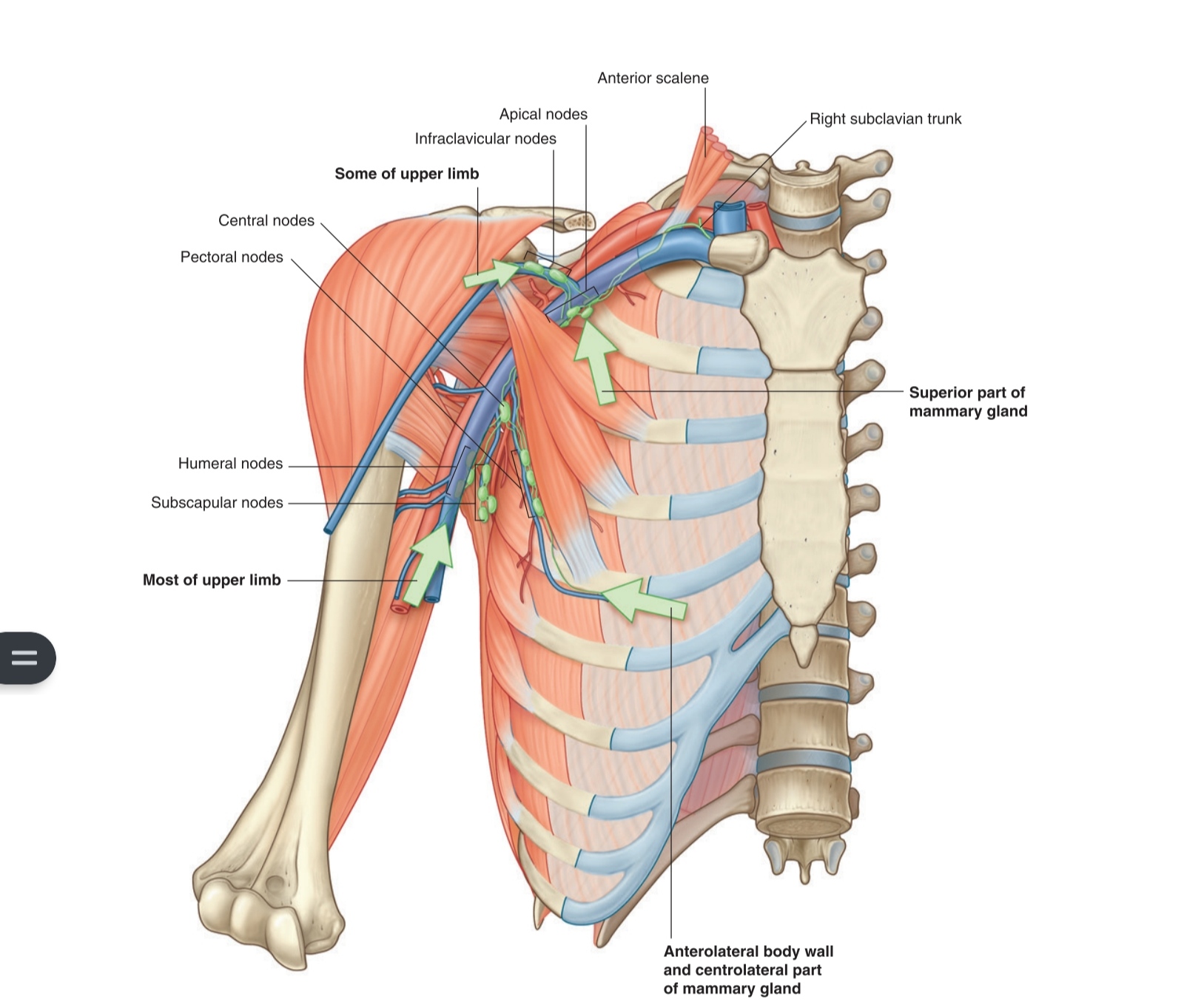
Axillary groups of lymph nodes
1_Lateral group: present in axilla along the axillary veen (medial side).
They receive afferent from the medial side of the upper limb (through
the supratrochlear lymph nodes).
They send efferent to the central and the apical groups.
2_ Anterior (pectoral) group: present along the lower border of the
pectoralis minor (along the lateral thoracic vessels).
They receive afferent from the front of trunk above the umbilicus and
the lateral quadrant of breast.
They send efferent to the central and the apical lymph nodes.
3_ Posterior (subscapular) group: present in the posterior wall of the
axilla along the subscapular vessels.
They receive afferent from the back of trunk above iliac crest.
They send efferent to the central and the apical lymph nodes.
4_Central group: present embedded in fat of axilla near its base.
They receive afferent from the anterior, the posterior and the lateral
groups.
They send efferent to the apical lymph nodes.
5. Apical group: present in apex of axilla, medial to the axillary v (under clavicle).
They receive afferent from all the other groups and the upper quadrant
of breast.
They send efferent by subclavian trunk to the thoracic duct in the left
side or right lymphatic duct in the right side.
They also send to the deep cervical lymph nodes.
Sources and References
1_DR.SAMEH DOSS, ANATOMY HAND_OUT,2015, (45_46).
2 Keith L. Moore, Arthur F. Dalley, Anne M.R. Agur,MOORE,seven edetion,2014.
picture:
1_MOORE,fig 7.38
2_MOORE,fig 7.37
3 MOORE,fig 7.40
4_MOORE,fig 7.47
5 MOORE,fig 7.57
6_MOORE,fig 7.58
2 Keith L. Moore, Arthur F. Dalley, Anne M.R. Agur,MOORE,seven edetion,2014.
picture:
1_MOORE,fig 7.38
2_MOORE,fig 7.37
3 MOORE,fig 7.40
4_MOORE,fig 7.47
5 MOORE,fig 7.57
6_MOORE,fig 7.58
