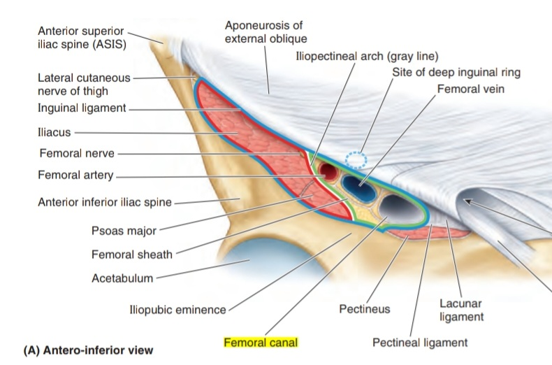
Femoral canal
By : Safa QusayInformation about femoral canal:
is the smallest space of the three compartments of the femoral sheath . It is conical in shape and short in long ( about 1.25-2 cm).
The space of the femoral canal is between:
1. Medially : the medial edge of the femoral sheath
2. Laterally : the femoral vein
The femoral canal extends to the level of the proximal edge of the saphenous opening to allow the femoral vein to expand when :
1. The venous return from the lower limb is increased ,
2. Or when the increased intra-abdominal pressure causes a blockage in the veins as during the Valsalva monenter
The space of the femoral canal is between:
1. Medially : the medial edge of the femoral sheath
2. Laterally : the femoral vein
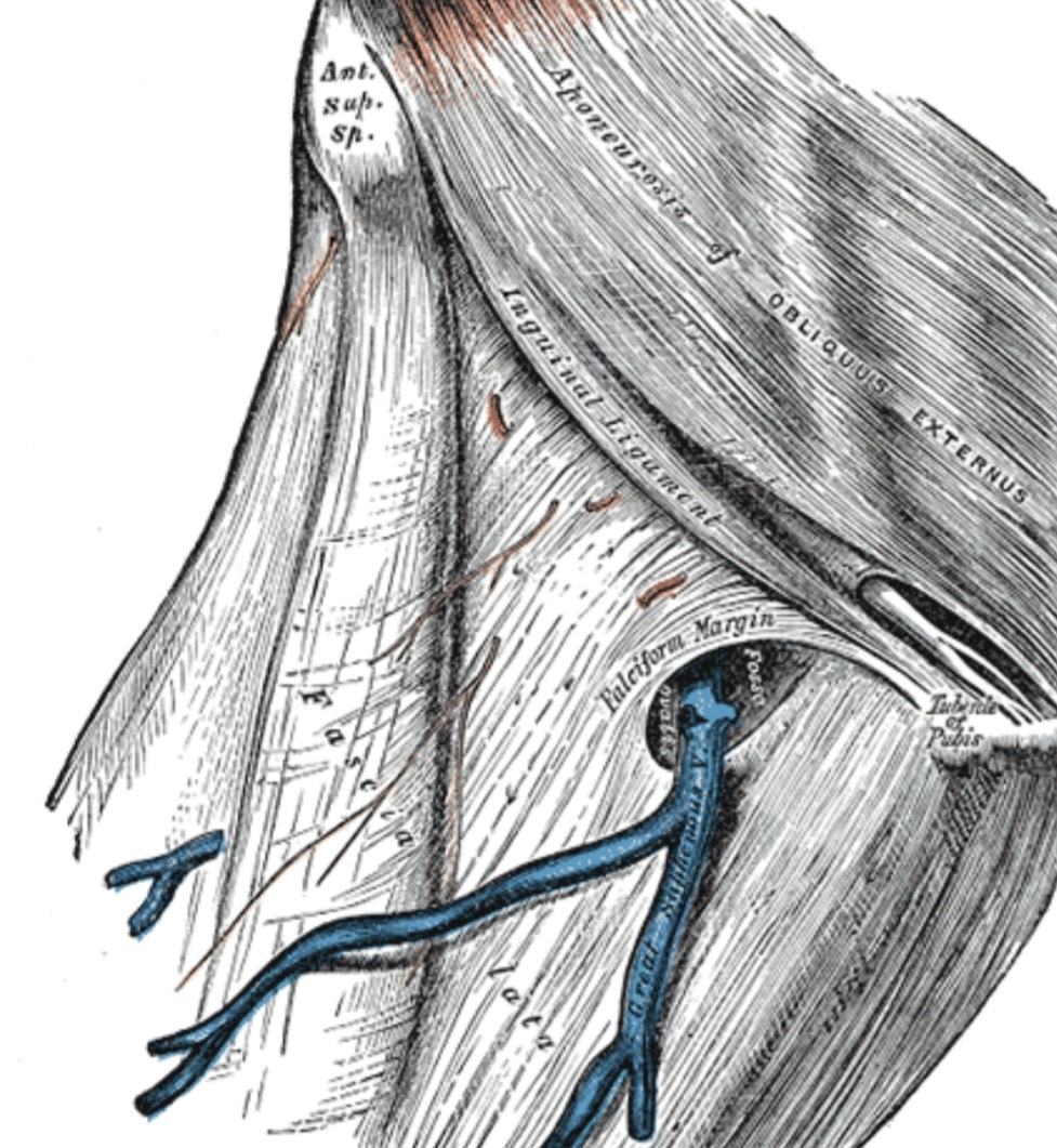
The femoral canal extends to the level of the proximal edge of the saphenous opening
Note
( saphenous hiatus , also fossa ovalis ) It is an oval opening in shape ,that is located in the mid of the upper part of the fascia lata of the thigh.1. The venous return from the lower limb is increased ,
2. Or when the increased intra-abdominal pressure causes a blockage in the veins as during the Valsalva monenter
Note
which means that the forceful expiration against a closed mouth and nose will increases the intrathoracic pressure ,it is has many benefits as in the pressure equalization of nose and sinuses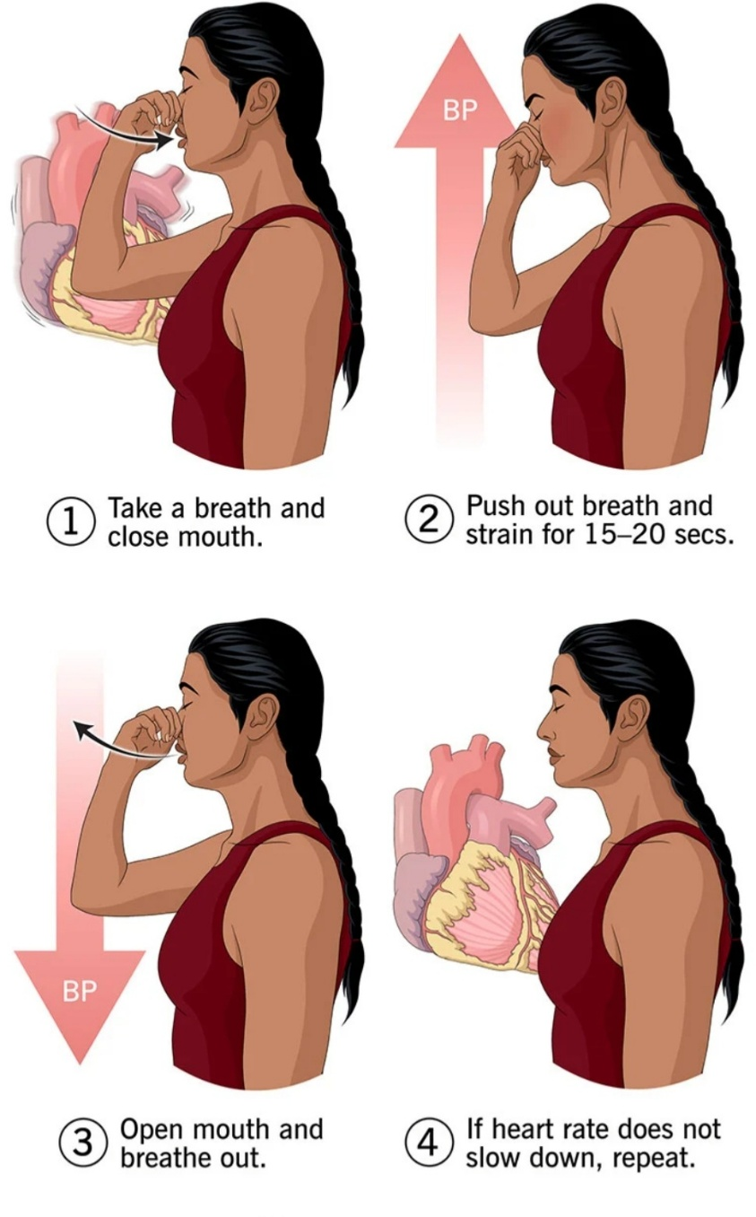
Valsalva monenter
The femoral canal Contains:
1. Loose connectives tissue
2. fat
3. An inguinal lymph node ( may be Cloquet node ).
4. Deep lymph node( lacunar node ).
2. fat
3. An inguinal lymph node ( may be Cloquet node
Note
It is a node of the inguinal lymph nodes types. We can found this node in femoral canal. Sometimes doctors are not sure if there is a hernia or enlargement of this node.4. Deep lymph node( lacunar node ).
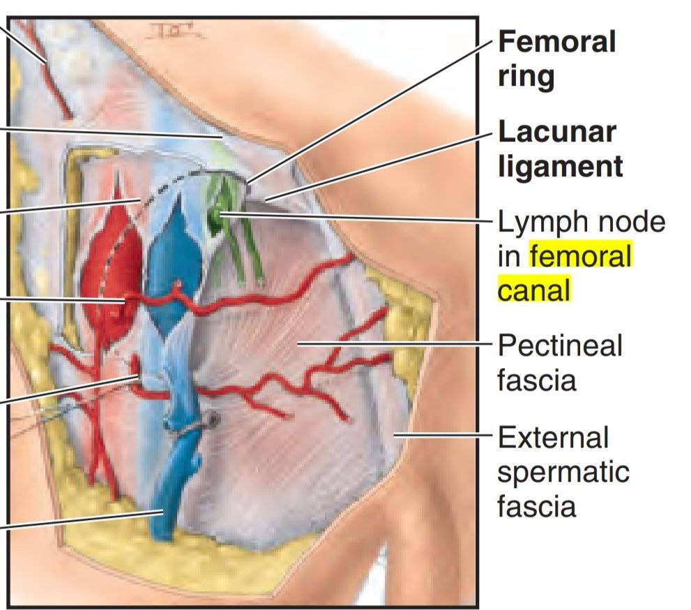
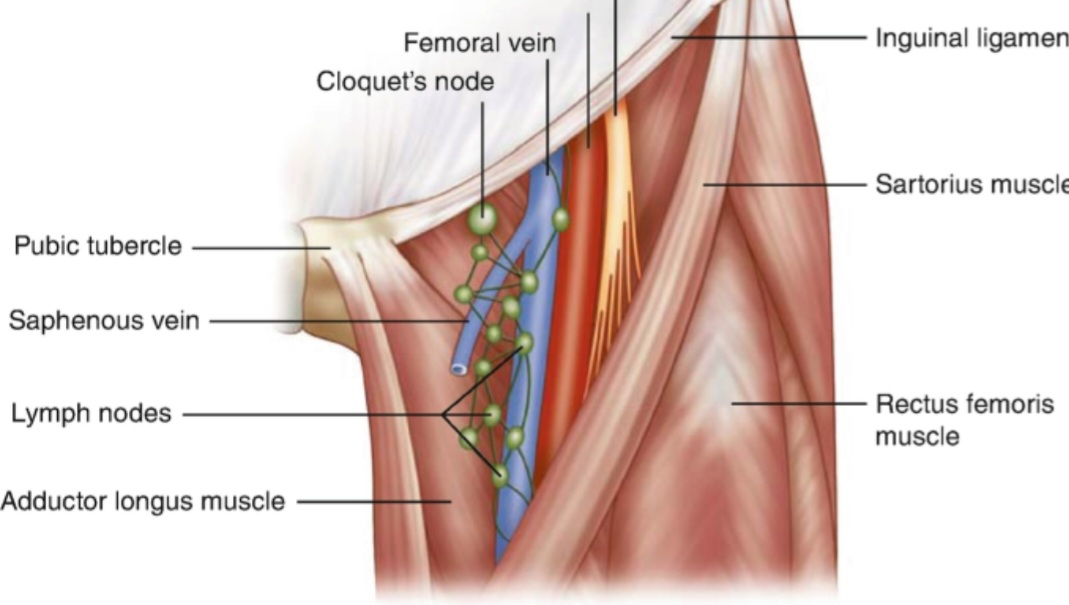
femoral canal Contains
Borders of the femoral canal:
1. Medial border: by the lacunar ligament
2. Lateral border: by the femoral vein .
3. Anterior border: by the inguinal ligament
4. Posterior border: by the:
• Pectineal ligament
• Superior ramus of the pubic bone
• The pectineus muscle
2. Lateral border: by the femoral vein .
3. Anterior border: by the inguinal ligament
4. Posterior border: by the:
• Pectineal ligament
• Superior ramus of the pubic bone
• The pectineus muscle
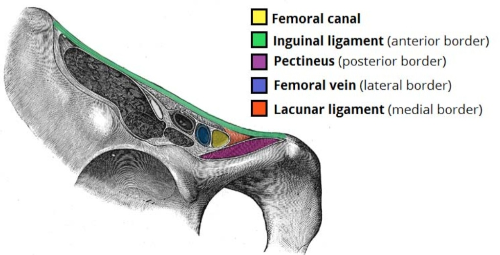
The The base of the femoral canal is the femoral ring (this ring is directed upward and it’s oval in shape ).
How this ring has been formed? by the small opening that we can found at its abdominal end .It is closed by extra peritoneal fatty tissue that forms the femoral septum.
How this ring has been formed? by the small opening that we can found at its abdominal end .It is closed by extra peritoneal fatty tissue that forms the femoral septum.
Note
A condensed portion of the fatty tissue that closes the femoral ring, named the septum femoral. The septum is pierced by lymphatic vessels for connecting the inguinal node and external iliac lymph nodes .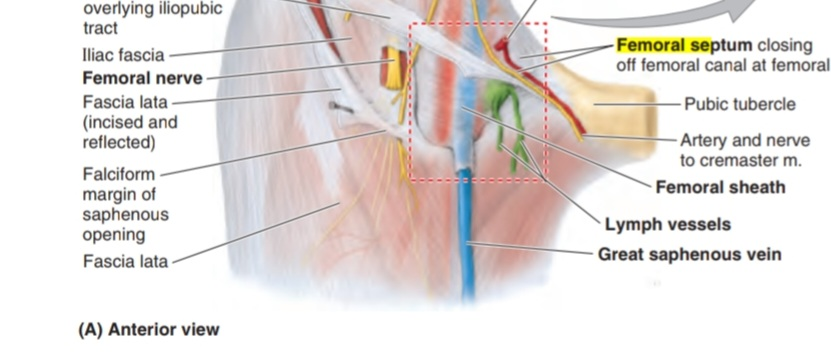
Femoral ring
The entrance of the femoral canal is the femoral ring (which means that the canal entrance opening in its superior border called femoral ring ).
Femoral ring contents are Cloquet's node and lymphatics. The femoral hernia mainly occurs in this ring because this area is so weak .
Femoral ring contents are Cloquet's node and lymphatics. The femoral hernia mainly occurs in this ring because this area is so weak .
The borders of the femoral ring:
1. Anteriorly : by the inguinal ligament
2. Medially : by the lacunar ligament
3. Laterally : by the medial side of femoral vein
4. Posteriorly :by the pectineal ligament and pectineus muscle
2. Medially : by the lacunar ligament
3. Laterally : by the medial side of femoral vein
4. Posteriorly :by the pectineal ligament and pectineus muscle
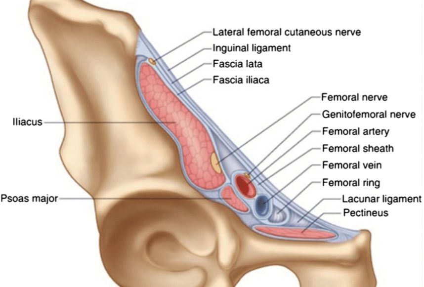
borders of the femoral ring
Clinical notes about (femoral canal)
Hernia
We can found many types of hernia are occurring in different parts of the body. Represent groin area, the lower part of the muscle wall of abdomen, and when the groin hernias pokes through this part, the hernia develops. Hernia happens if the tissue passes through a weak spot that is found in muscle wall of the groin.
Common causes of hernia include:
1. Obesity
2. Overstraining while coughing.
3. Exercising.
4. Having a low birth weight
5. Being older
6. A personal or family history of hernias
Common causes of hernia include:
1. Obesity
2. Overstraining while coughing.
3. Exercising.
4. Having a low birth weight
5. Being older
6. A personal or family history of hernias
Types of the hernia
Hernia includes many types ,as:
• Femoral hernia
• Inguinal hernia
• Epigastric hernia
• Hiatal hernia
• Femoral hernia
• Inguinal hernia
• Epigastric hernia
• Hiatal hernia
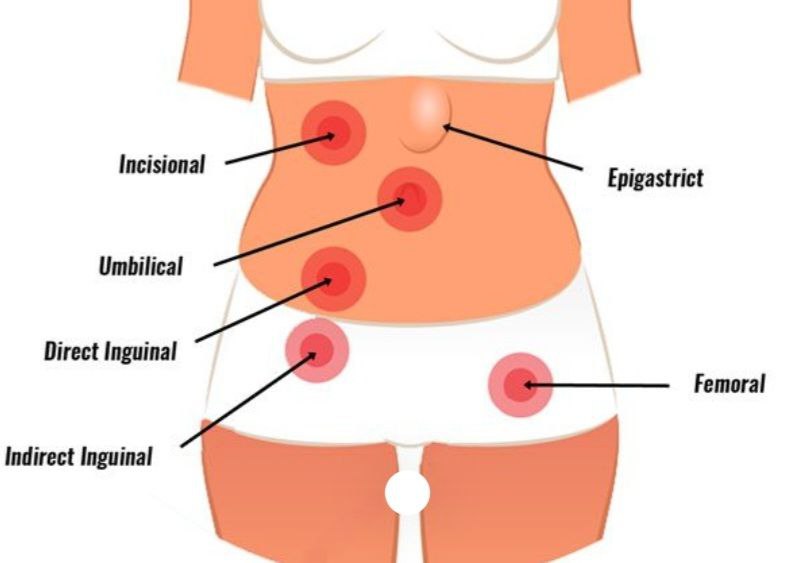
Types of hernias
First type femoral hernias
It occurs when tissue, such as part of the bowel, pokes through the groin’s muscle wall at the top of the inner thigh. It passes through a weak spot of the femoral canal because it was a weak area.
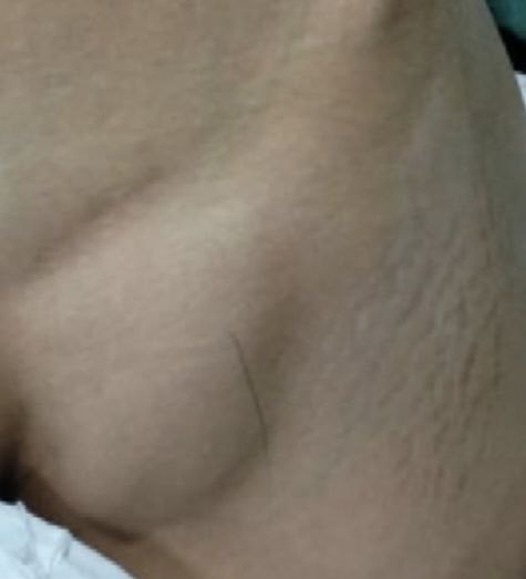
The femoral hernia includes 3 types :
1. Incarcerated femoral herni a( it called irreducible or trapped femoral hernia, this happens if the hernia becomes stack in the femoral canal and cannot return into the abdomen.
2. Obstructed femoral hernia (included the hernia and a section of the intestine becoming entangled),
3. Strangulated femoral hernia ( happen if the hernia prevents blood from reaching the bowel).
The femoral hernia happen more often in female and particularly in those who are older. Why ?
Because the wider structure of female pelvis.
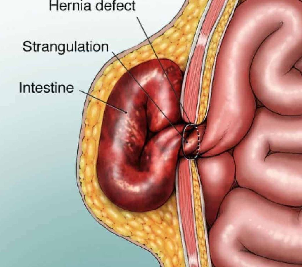
Strangulated femoral hernia
The symptoms
1. Groin discomfort that may get worse during standing, lifting, or straining
2. Abdominal pain
3. Nausea
4. Vomiting
5. Lump on the upper inner thigh or groin may be painful
The Diagnosis
Doctor examines the area, and asks you to do imaging tests, such as ultrasound, CT, or MRI scans..
1. Groin discomfort that may get worse during standing, lifting, or straining
2. Abdominal pain
3. Nausea
4. Vomiting
5. Lump on the upper inner thigh or groin may be painful
The Diagnosis
Doctor examines the area, and asks you to do imaging tests, such as ultrasound, CT, or MRI scans..
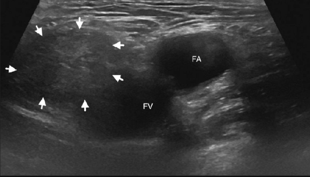
Femoral hernia (x-ray picture)
The treatment:
Surgical repair is necessary, because femoral hernias can cause severe complications. The surgery can be done by two manners depending on the:
1.Size of the hernia
2.Person’s age
3. Person’s general health and other factors
Types of Surgical hernia :
1. Laparoscopic
2. Open
Surgical repair is necessary, because femoral hernias can cause severe complications. The surgery can be done by two manners depending on the:
1.Size of the hernia
2.Person’s age
3. Person’s general health and other factors
Types of Surgical hernia :
1. Laparoscopic
2. Open
Second types of hernias (inguinal hernia)
The most common type of hernia is the inguinal hernia Inguinal hernias happen when a part of the membrane lining the abdominal cavity intestine enters the weak spot that is found in the abdomen - often along the inguinal canal which carries the spermatic cord in males . In women, the inguinal canal carries a ligament that is helps to hold the uterus in place. Watch out, for this reason it is more common in males.
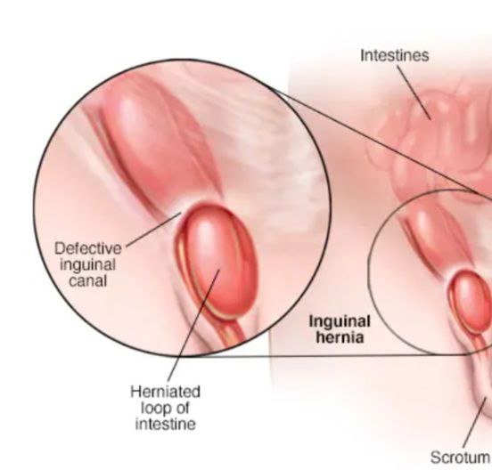
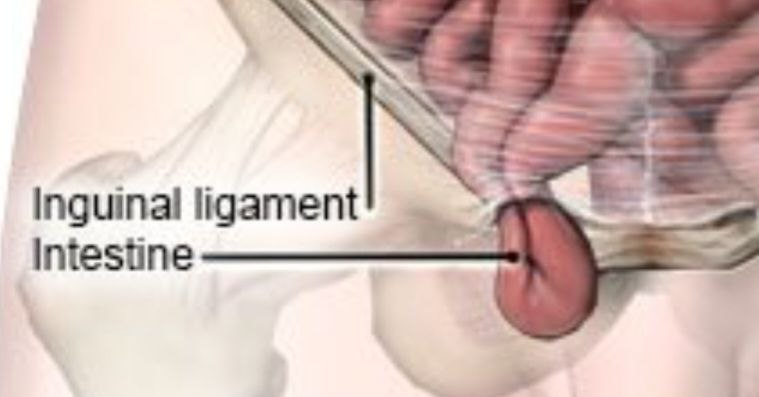
Inguinal hernia
Diagnosis
Physical exam is often all what's needed to diagnose an inguinal hernia.
The doctor will look a bulge in thigh
If the diagnosis isn't readily apparent, The doctor might ask to do the imaging test, such as an abdominal ultrasound.
Treatment
If the hernia is small and isn't bothering , the doctor might recommend watchful waiting. There is some ways to treat as wearing a supportive truss may help relieve symptoms,
Enlarging or painful hernias often require surgery.
There are two general types of hernia operations
1. Open hernia repair
2. Minimally invasive hernia repair.
Symptoms
Include:
• A burning or aching sensation at the bulge
• Pain or discomfort in the groin, especially when bending over, coughing or lifting
• A heavy or dragging sensation in the groin
• Weakness
• Pressure in the groin
References:
1.The femoral canal, Oliver Jones, teacheme anatomy https://teachmeanatomy.info/lower-limb/areas/femoral-canal/
2.Figure of the femoral canal border from teacheme anatomy
3.Figure of cloquet node from springer
4.Figure of saphenous opening from Wikipedia
5. Figure of theValsalva Maneuvers from Cleveland Clinic
6.Femoral Hernia, by Elaine K. Luo ,M.D. ,healthline
.https://www.healthline.com/health/femoral-hernia
7.MOORE Clinically Oriented ANATOMY Keith L. Moore Arthur F. Dalley Anne M.R. Agur(532,202_203)
8.Recurrent Hernia : Prevention and Treatment ,Robert J. Fitzgibbons Volker Schumpelickall 365,360
9.Moore Clinically Oriented ANATOMY Keith L. Moore Arthur F. Dalley Anne M.R Figure 5.27.and Figure 5.28. A,b
