Humerus
By : Sara OthmanOverview
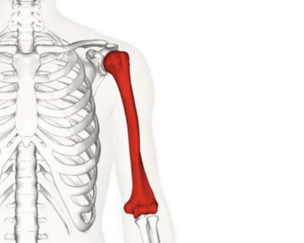
The humerus is the largest and longest bone of the upper limbs
Location:- is the only bone in the arm.
Type:- long bone ( has shaft and two epiphysis ).
*shaft is the body of the bone that composed of compact bone which surrounds a bone marrow cavity which contains red or yellow marrow , the shaft is the first part of the bone that undergoes ossification.
*epiphysis is the end of the bone which is made of spongy bone and covered by compact bone , it is filled with red bone marrow which produces red bone cells ( erythrocytes ) , the epiphysis of the humerus is classified as the pressure epiphysis and forms part of the shoulder joint.
Bony Parts
1- Its proximal end is the rounded smooth head of humerus which make shoulder joint ( glenohumeral joint ) with glenoid cavity of scapula.
2- the anatomical neck is surrounding the articular surface of the head and located inferior to the head and medial to the greater tubercle .
3- It has two tubercle ( lesser and greater ) which are important for many attachments of the muscles
A) on greater tubercle ( insertion of supraspinatus m. , infraspinatus m. , teres minor m. )
B) on lesser tubercle ( insertion of subscapularis m.
4- below the tubercles there is a surgical neck which wind around it the axillary n. and the posterior and anterior circumflex humeral vessels ( from the 3rd part of axillary a. ) [on other source, they only mention the posterior circumflex humeral a. and the axillary n.
5- bicipital groove ( inter-tubercular groove ) is a deep groove on the anterior surface of humerus between the lesser and greater tubercles which is occupied by the tendon of the long head of the biceps. and is important for many attachments of the muscles ( latissimus doris m. , pectoralis major m. , teres major m. )
6- the oblique spiral or radial groove for ( redial n. and profunda brachii a. ) at the back of humerus.
7- at the lateral surface of the middle third of the humerus there is a deltoid tuberosity ( the site of insertion of deltoid m. )
— at the distal third there are
8- two epicondyle (medial and lateral) they are subcutaneous.
A) the medial one is more prominent than the lateral part , the medial epicondyle gives origin to the flexor muscles of forearm, behind medial epicondyle the ulnar n. is passed in the ulnar sulcus and the fracture of medial epicondyle will hurt the ulnar n.
B) the lateral one gives origin to extensor muscles of forearm.
9- it has two supracondylar ridges (medial and lateral) just proximal to condyles.
10- there are three fossae at the lower end, one posterior and two anterior
* posteriorly there is an olecranon fossa ( triangular depression ) for olecranon process of ulna and is deeper than the anterior fossae.
* anteriorly there are coronoid fossa for coronoid process of ulna and radial fossa for the head of radius.
11- there are capitulum ( articulation of radius ) and trochlea ( medial portion of
articular surface ).
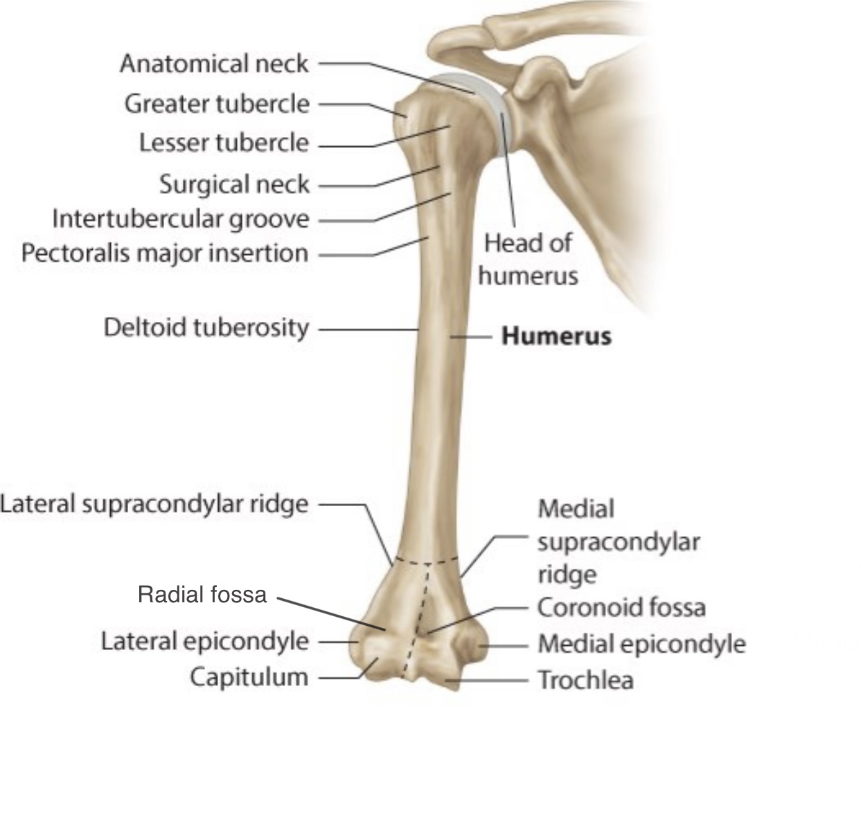

Anterior view of right humerus
How to know the bone is right or left
1- the head ( proximal end ) should be directed anteriorly and medially.
2- the olecranon fossa ( large fossa ) should be posteriorly.
3- intertubercular groove should be forwarded and upwarded.
4- medial epicondyle is more prominent than the lateral .
2- the olecranon fossa ( large fossa ) should be posteriorly.
3- intertubercular groove should be forwarded and upwarded.
4- medial epicondyle is more prominent than the lateral .
The articulaion
the humeral bone makes two joint with the other bones:
1- Proximately, the shoulder joint : its type is synovial ball and socket , it forms between the head of humerus ( the ball ) and the glenoid cavity of scapula ( the socket ).
2- Distally, the elbow joint which forms between the trochlea and capitulum of humerus and trochlear notch of ulna and head of radius , its type is synovial hinge joint.
* synovial joints are joints that have a cavity between the bones filled with synovial fluid.
* ball and socket joint allows the movement in many directions
* hinge joint allows bones to move in one direction back and forth with a limit motion along other planes.
Muscles attached to the bone
The humerus gives attachment to many important muscles ,
it receives the insertion of
1- Rotator cuff m. ( subscapularis , infraspinatus , supraspinatus , teres minor ) at the proximal end ( greater & lesser tubercles ).
2- The major m. ( pectoralis major , teres major ) at the lips of intertubercular groove.
3- Latissimus dorsi at intertubercular groove.
4- Deltoid at deltoid tuberosity.
5- Coracobrachialis at medial surface of middle third of humerus.
And gives origin to
1- Lateral & Medial head of triceps brachii at the posterior surface of humerus.
2- Brachialis at anterior surface of humerus.
3- Brachioradialis at lateral supracondylar ridge.
4- Anconeus & extensor carpi radialis longus at lateral epicondyle.
5- Pronator teres and flexor m. ( flexor carpi radialis , flexor carpi ulnaris , flexor digitorum superficialis , palmaris longus ) at medial epicondyle.
6- Supinator & extensor m. ( extensor carpi radialis brevis , extensor carpi ulnaris , extensor digiti minimi , extensor digitorum ).
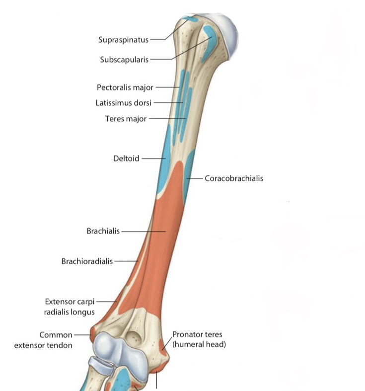
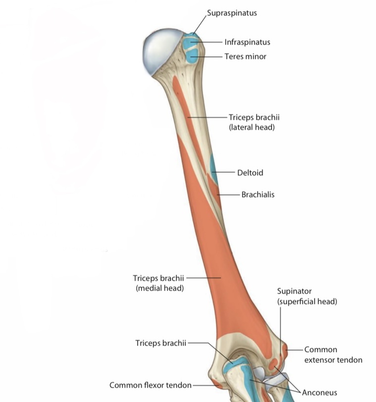
Attachments on anterior surface
The structures related to the humerus
1- axillary n. and posterior circumflex humeral a. around surgical neck.
2- redial n. and profunda brachii a. in spiral groove.
3- the ulnar n. behind medial epicondyle.
4- the median n. in front of distal third of humerus
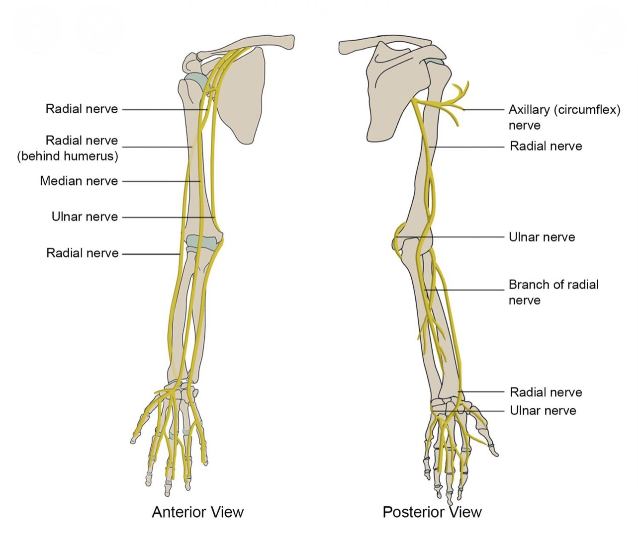
Nerves engaged to the humerus
Fractures of humerus
A) The fracture at the surgical neck is the most common fracture in the proximal end , it can be caused by a minor fall on the hand or from directed blow to the area and that can hurt the posterior circumflex humeral a. and the axillary n. which may cause the paralysis of deltoid and teres minor musles that leads to loss of abduction from 15-90 degrees and causes flat shoulder and loss the sensation of the skin over the distal half of deltoid m.
B) The fracture could be horizontally at the mid-shaft in the spiral groove and hurts the radial n. that leads to wrist drop and finger drop because the paralysis of muscles of back of forearm and part of triceps muscle ( not completely ) and loss of sensation along all cutaneous branches of radial n. except posterior cutaneous n. of arm ( which is given in axilla ) as well as the profunda brachial a. can be hurt too.
C) the fracture of medial epicondyle where the place of the ulnar n. will lead to partial claw hand ( paralysis of flexor carpi ulnaris m. , medial half of flexor digitorum profundus m. and hand muscles ) and loss of sensation of medial third of hand ( palmar & dorsal ) and skin of the medial one and half fingers ( palmar & dorsal ).
D) the fracture between two epicondyles can be caused by a severe fall on the flexed elbow and that damages the median n. and cause Ape-like deformity ( loss of flexion & opposition of thumb ), loss pronation of forearm and flexion of proximal and distal phalanges of medial four fingers and flexion of distal phalanges of index and middle fingers and loss of sensation of lateral two thirds of palm of hand and skin of lateral three and half fingers.

Ftactures of humerus
Blood supply of humerus
It receive its blood supply from the branch of the brachial a. which pierces the middle third at the insertion of coracobrachialis m. , the head is supplied mainly by anterior circumflex humeral a. where the anterolateral a. ( branch of it ) and arcuate a. is continuation of ascending branch of it perice the head at intertubercular groove as well as supply the greater and lesser tubercles.
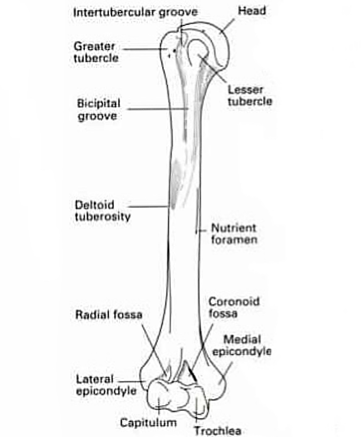
Nutrient foramen of the humerus
Anastomosis around the humerus
Anastomosis around surgical neck
1-anterior circumflex humeral a. ( from the 3rd part if axillary a. )
2- posterior circumflex humeral a. ( from the 3rd part of axillary a. )
3- ascending branch of profunda brachii a.
Anastomosis around elbow
A) In front of medial epicondyle :
1- Anterior branch of inferior ulnar collateral a. ( from brachial a. )
2- Anterior ulnar recurrent a. (from ulna a.)
B) Behind medial epicondyle :
1- Superior ulnar collateral a. & posterior branch of inferior ulnar collateral a.
2- Posterior ulnar recurrent a. (from ulnar a.)
C) In front of lateral epicondyle :
1- Anterior descending branch of profunda brachii a. (from brachial a.)
2- Radial recurrent a. (from radial a.)
D) Behind lateral epicondyle :
1- Posterior descending branch of profunda brachii a. (from brachial a.)
2- Interosseous recurrent a. ( from ulnar a.)


Anastomosis around surgical neck of humerus
References
1- Moore's Clinically Oriented Anatomy 7th Edition / pp.676-677 , pp.684-685
2- LAWRENCE E. WINESKI Snell's Clinical Anatomy by Regions /10th Edition (2018 ) / pp.90-91-92-93-94
3- Humerus , Author: Charlotte O’Leary BSC, MBChB , Source: Kenhub , https://www.kenhub.com/en/library/anatomy/the-humerus
4- The humerus , Author: Oliver Jones , Source: TeachMe Anatomy , https://teachmeanatomy.info/upper-limb/bones/humerus/
5- Blood supply of the humerus , Wheeless’ Textbook of Orthopaedics , https://www.wheelessonline.com/joints/blood-supply-to-the-humerus/
References of images
_Cover image , the Humerus , source: TeachMe Anatomy , https://images.app.goo.gl/ocCdAjpse7f1nWHx8
_figure 1 & 2 , anterior & Posterior view of humerus , source: Black Shirks , https://images.app.goo.gl/rA3iT4pN6hNVoqRn8
_figure 3 & 4, attachments , Gray’s atlas of anatomy / plate407
_figure 5 , Nerves engaged to the humerus , Source: Health house Clinics , https://images.app.goo.gl/wBYDQmTE3MNacKkx8
_figure 6 , Fractures of humerus , Source: Anatomy QA , https://images.app.goo.gl/Da683k6WZ89S5A7v5
_ figure 7 , Nutreint foramen of he humerus , Source: Black Shirts , https://images.app.goo.gl/rgN7hu4fXAPxJc1x6
_figure 8 , Anastomosis around surgecal neck , source: orthopeadics and trauma , https://images.app.goo.gl/vWUNtxuRotgfzsES7
_figure 9 , Anastomosis around the elbow , Source: Quizlet ، https://images.app.goo.gl/UhzNHaQiLjZ8YFKk7
