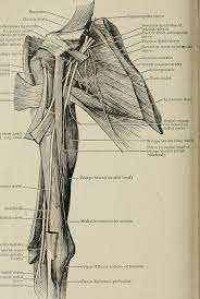
Triceps Brachii (TB)
By : Fahad Al-BakNaming: -
The naming: -
/TRY-seps BRAY-key-eye/
From the name we can understand that it consists of three heads [tri- meaning three], also we get that it’s located over the arm [brachium means the arm].
/TRY-seps BRAY-key-eye/
From the name we can understand that it consists of three heads [tri- meaning three], also we get that it’s located over the arm [brachium means the arm].
General: -
-The Triceps Brachii is the muscle of the posterior compartment of the arm, it is the only muscle in the posterior part of the arm; it is a large fusiform muscle; spanning almost the entire length of the humerus, It belongs to the Muscles of the Elbow and Radioulnar Joints as while.
It is located the lateral intermuscular septum separates the dorsal part from the arm from the ventral part, which is where the flexors of the arm are (biceps, brachialis, and brachioradialis).
The flexor muscles of the anterior compartment are almost twice as strong as the extensors in all positions; consequently, we are better pullers than pushers. It should be noted, however, that the extensors of the elbow are particularly important for raising oneself out of a chair, and for wheelchair activity. Therefore, conditioning of the triceps is of particular importance in elderly or disabled persons.
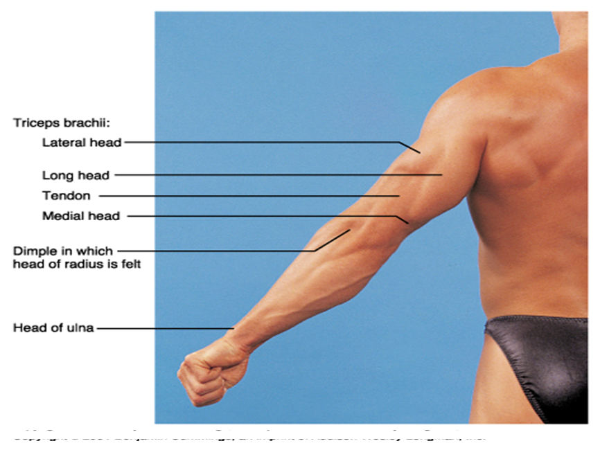
-The TB have three heads, their names are:
The Lateral head (on the lateral side of the arm).
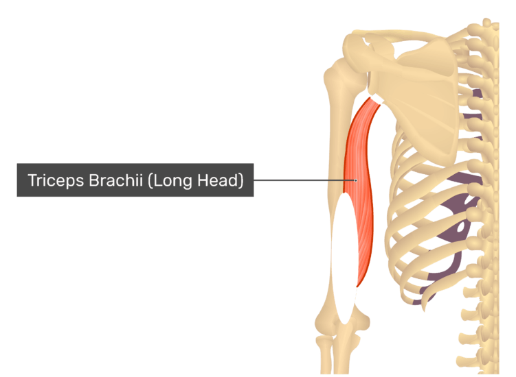
The Long head (the middle one).
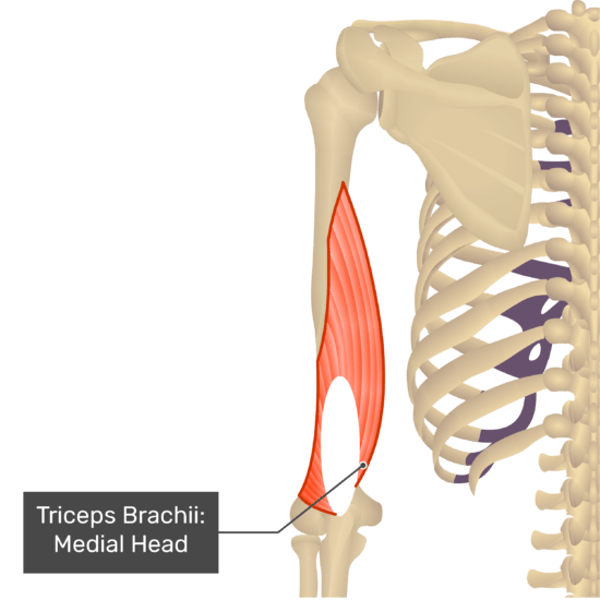
The Medial head (on the medial side of the arm).
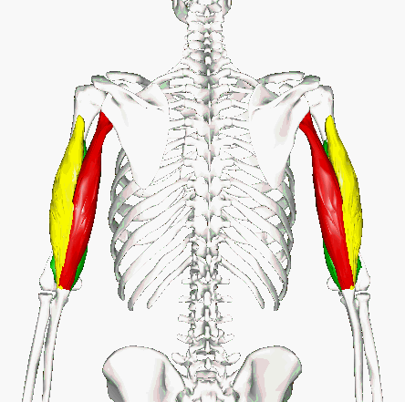
Surface: -
- the triceps can be visible on the posterior side of the whole arm.
The Agonist & Antagonist: -
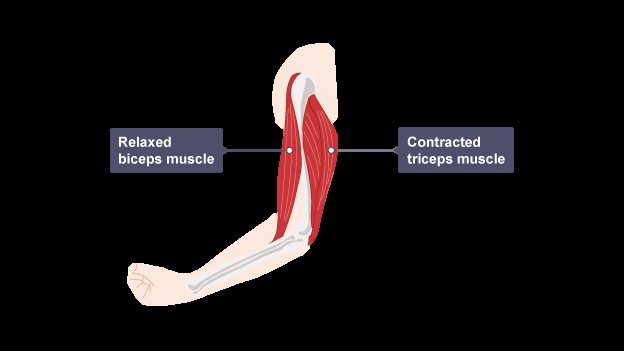
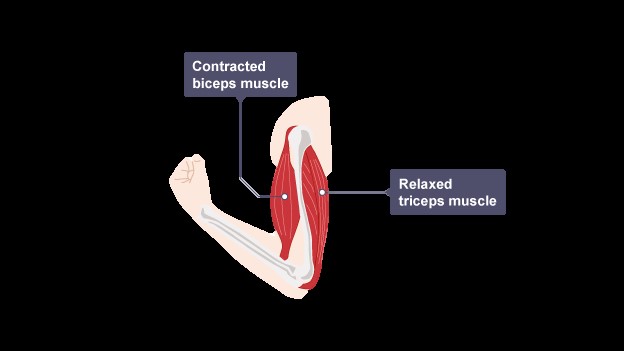
In an antagonistic muscle pair as one muscle contracts the other muscle relaxes or lengthens. The muscle that is contracting is called the agonist and the muscle that is relaxing or lengthening is called the antagonist.
One way to remember which muscle is the agonist – it's the one that's in 'agony' when you are doing the movement as it is the one that is doing all the work.
The Biceps Brachii and Triceps Brachii are agonist and antagonist, where when the Biceps Brachii is flexed the Triceps Brachii is relaxed and vice versa
Supply: -
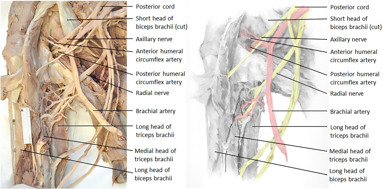
-The blood supply comes from the Deep Brachial Artery (a branch of the Brachial Artery) and the Circumflex Scapular Artery (a branch of the Subscapular Artery).
-The Triceps Brachii has a nerve supply from Radial n.
Origin & Insertion: -
The triceps brachii is located in the dorsal compartment of the arm. The lateral intermuscular septum separates the dorsal part from the arm from the ventral part, which is where the flexors of the arm are (biceps, brachialis, and brachioradialis).
Origins (The immoveable part):
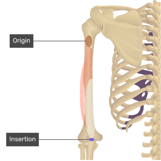
Two of the heads arise from the humerus, being separated by the spiral groove, and the third from the scapula; the third head being the long head. The muscle is inserted to the olecranon of the ulna via a tendon known as the triceps tendon.
- The long head (infra-glenoid tubercle of the scapula): The long head of the triceps originate from infraglenoid tubercle of the scapula and the adjacent glenoid labrum and it blends in the shoulder capsule, the long head run decent with the medial head deep to it. While the long head is going downward from infraglenoid tubercle it goes anteriorly to teres minor and posteriorly to teres major and on it ways it for the medial side of the quadrangular space and the lateral side of the triangular space.
- The medial head (Posterior Shaft of the humerus; the distal 2/3 to the Olecranon Process of the Ulna): it arises from posterior side of the humerus medially and inferiorly to the radial groove and medial to it; there is an extra attachment to the posterior side of the lateral and medial intermuscular septa. The medial head lies deep to the others two heads.
- Lateral head (Posterior Shaft of the Humerus the proximal 1/3): it originates from the humerus in its posterior compartment on the lateral side of the arm (as the name suggested); it is to the out most of the posterior of the arm, it arises from the upper most of the humerus. It is above and lateral to the spiral groove; as the fibers pass to join with those of the medial head, they cover the spiral groove. The origin of the lateral head lays in-between the insertion of the Teres Major and Deltoid.
Insertion (moveable part):
The three heads of triceps come together to form a broad, laminated tendon, the superficial part of which covers the posterior aspect of the lower third of the muscle, while the deeper part arises from within the substance of the muscle. This arrangement provides a larger surface area for the attachment of the muscle fibers. Both laminae blend to form a single tendon which attaches to the posterior part of the proximal surface of the olecranon of the ulna, and to the deep fascia of the forearm on either side. Some muscle fibers from the medial head attach to the posterior part of the capsule of the elbow joint serving to pull it clear of the moving bones preventing it becoming trapped during extension of the joint.
The three heads of triceps come together to form a broad, laminated tendon, the superficial part of which covers the posterior aspect of the lower third of the muscle, while the deeper part arises from within the substance of the muscle. This arrangement provides a larger surface area for the attachment of the muscle fibers. Both laminae blend to form a single tendon which attaches to the posterior part of the proximal surface of the olecranon of the ulna, and to the deep fascia of the forearm on either side. Some muscle fibers from the medial head attach to the posterior part of the capsule of the elbow joint serving to pull it clear of the moving bones preventing it becoming trapped during extension of the joint.
Morphology (four headed triceps brachii):
There have been reports of a fourth head in a few cadaver studies. This accessory muscle can originate from the humerus, shoulder capsule, coracoid processes, or muscles in close proximity. These accessory muscles can potentially compress the radial and ulnar nerves. Therefore, it is essential for surgeons and physicians to keep in mind that although rare, these variations exist, which can help diagnose cases of nerve entrapment and other pathologic causes that may not be explained by any other typical factors.
There have been reports of a fourth head in a few cadaver studies. This accessory muscle can originate from the humerus, shoulder capsule, coracoid processes, or muscles in close proximity. These accessory muscles can potentially compress the radial and ulnar nerves. Therefore, it is essential for surgeons and physicians to keep in mind that although rare, these variations exist, which can help diagnose cases of nerve entrapment and other pathologic causes that may not be explained by any other typical factors.
Motion & Action: -
( Extension of the forearm, extension of the arm, adduction of the arm and horizontal extension of the arm )
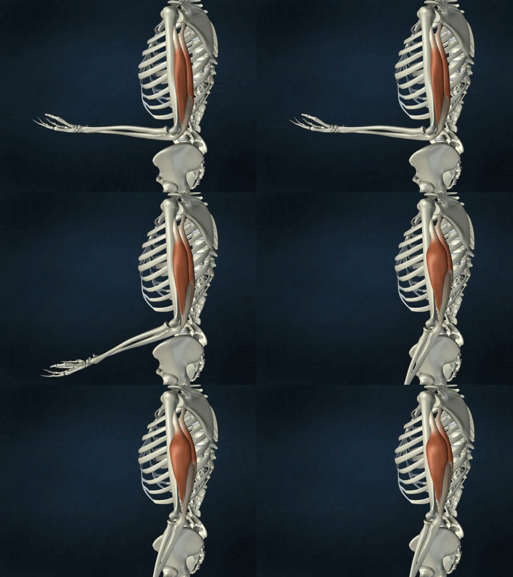
The main action for the triceps brachii is to extend the forearm at the elbow joint, while the anterior compartment of the arm is the responsible for flexing the forearm; gravity by itself can take care of extending the forearm however the triceps come in place when the speed of the extension matters as in punching. The triceps is a great pusher and is what we use to get out of chairs for example.
At rest the arm is slightly flexed due to the Biceps brachii over powering the triceps.
Along with extending the forearm at the elbow joint, the triceps can also stabilize the elbow joint when the forearm and hand are doing fine movements such as writing.
Triceps brachii is also an important extensile ‘ligament’ on the undersurface of the shoulder joint capsule during abduction of the arm.
All of the triceps head function whether the arm is supinated or pronated with lateral head being the strongest in between the three.
Aside to that; the long head of the triceps actives other tasks, Because it attaches to the scapula, the long head not only extends the elbow but will also have a small action on the glenohumeral or shoulder joint. With the arm adducted, the triceps muscle acts to hold the head of the humerus in the glenoid cavity. This action helps prevent any displacement of the humerus. The long head also assists with the extension and adduction of the arm at the shoulder joint.
In The Field Notes: -
-Testing The Triceps Brachii: - (or to determine the level of a radial nerve lesion), the arm is abducted 90° and then the flexed forearm is extended against resistance provided by the examiner. If acting normally, the triceps can be seen and palpated. Its strength should be comparable with the contralateral muscle, given consideration for lateral dominance (right or left handedness).
-Land mark: the triceps brachii also plays a role in creating anatomical spaces which are traversed by neurovascular structures. This makes the triceps brachii muscle an important surgical landmark.
-Land mark: the triceps brachii also plays a role in creating anatomical spaces which are traversed by neurovascular structures. This makes the triceps brachii muscle an important surgical landmark.
Clinical Note: -
• Triceps Brachii Tendon Injuries: - Distal triceps ruptures are rare injuries due to the special anatomical features of the muscle and tendon–bone junction.
• This injury typically occurs at the tendon–bone junction due to an eccentric contraction of the muscle.
• The treatment is controversial, especially in partial ruptures; surgical repair is indicated for complete ruptures of the distal triceps tendon.
• Several repair techniques have been described for acute complete ruptures.
• Chronic ruptures often require reconstruction rather than direct repair.
Relations: -
The triceps brachii is superficial in the posterior arm except for the proximal attachments of the long head and the lateral head, which are deep to the deltoid.
On the lateral side, the triceps brachii borders the brachialis proximally and the brachioradialis and extensor carpi radialis longus distally.
On the medial side, the triceps brachii borders the coracobrachialis and the brachialis.
The long head of the triceps brachii runs between the teres minor and the teres major.
The triceps brachii is located within the deep back arm line myofascial meridian.
Sources: -
- Moore Clinically Oriented Anatomy 7th Edition (735).
- Joseph E Muscolino - The Muscular System Manual: The Skeletal Muscles of the Human Body
Book (160-162).
- Anatomy, Shoulder and Upper Limb, Triceps Muscle – NLM
- Distal triceps ruptures – NLM
- Triceps brachii muscle – KenHub
- Joseph E Muscolino - The Muscular System Manual: The Skeletal Muscles of the Human Body
Book (160-162).
- Anatomy, Shoulder and Upper Limb, Triceps Muscle – NLM
- Distal triceps ruptures – NLM
- Triceps brachii muscle – KenHub
