


Elbow Joint
By : Yaser MohammedLocation and Articulation
Elbow joint: is a joint found in the upper limb, it works as a connection area between the arm and the forearm.
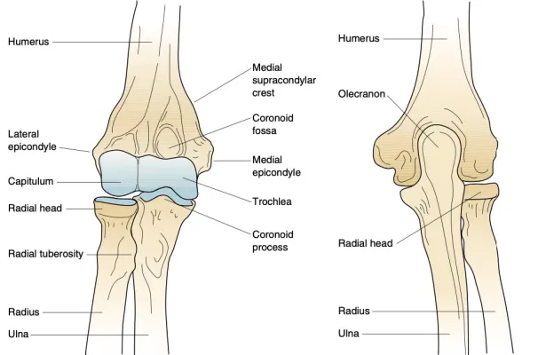
The elbow joint formed from the articulation of three bones: Humerus superiorly from the arm, Ulna medially, and Radius laterally, inferiorly from the forearm. these bones give rise to two joints:
1- Humeroulnar joint medially: The joint between the trochlea at the distal end of the humerus and the trochlear notch on the proximal ulna.
2- Humeroradial joint laterally: The joint between the capitulum on the distal end of the humerus with the head of the radius (proximal end).
The Humeroradial joint and Humeroulnar joint give the elbow joint hinge properties, due to rounded surfaces of the trochlea and capitulum and the concave surface of the trochlear notch of the ulna and head of the radius. The articular surfaces of these bones are separated from each other by a layer of hyaline cartilage .
At the elbow joint, other joints are formed between the proximal ends of the radius and ulna which articulate with each other at the proximal radioulnar joint . which is a synovial pivot joint.
.jpg)
Bony anatomy of the joints of the elbow—anterior view.
Joint Type and Its Movement
The elbow joint is a synovial hinge joint. uniaxial (move in only one plane), it has only two movement act on the forearm which are:
Flexion: (Range of motion from 0 to140 degrees) Act on decreasing the angle between the arm and forearm, the muscles responsible for this action are Biceps brachii and Brachialis , both innervated by the musculocutaneous nerve.
Extension: (ROM from the range of flexion to 0 degree for standard elbow) Act on increasing the angle between the arm and forearm, the muscle responsible for this action is the Triceps brachii innervated by the radial nerve.
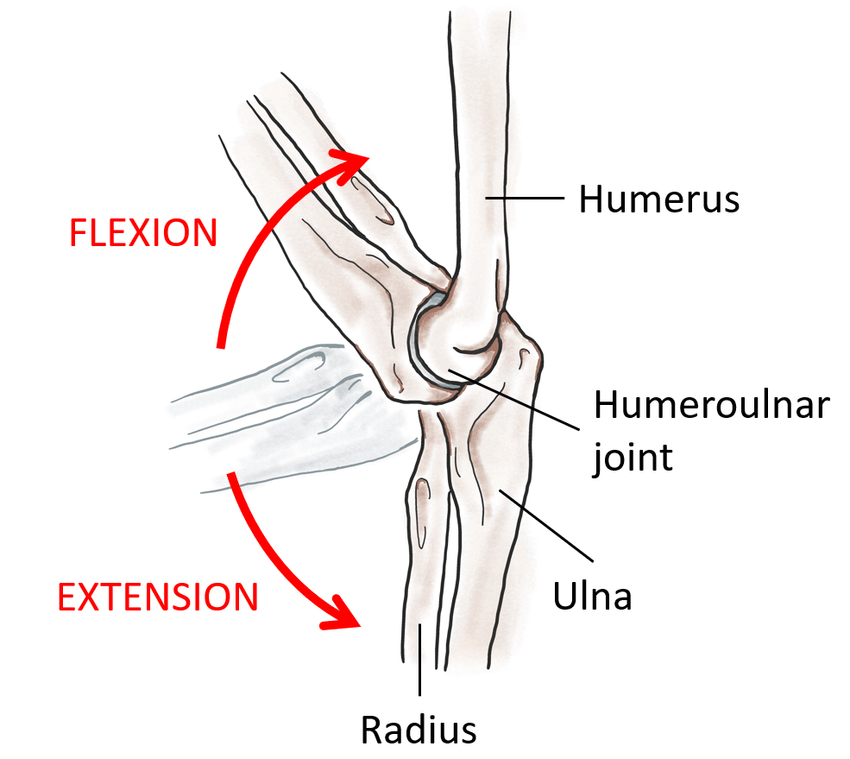
Elbow Joint Movements
Carrying Angle
The carrying angle: Is an angle made by the median axis of the arm and median axis of the forearm at full extension and supination. This angle permits the forearms to swing movements during walking and is essential when carrying objects. The long axis of the fully extended ulna makes an angle of approximately 170° with the long axis of the humerus. This means that the forearm and the hand should be 5 to 15 degrees away from the body, but in females, the carrying angle is slightly greater, Due to secondary sexual characteristics .
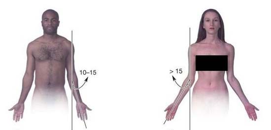
Carrying angle between males and females
Blood Supply
The Blood supply of the elbow joint is formed from anastomosing of many branches of different arteries, the Proximal part of the elbow joint is supplied by the superior and inferior ulnar collateral artery (branches from brachial artery), radial collateral artery, and middle collateral artery (branches from profunda brachii).
While the Distal part of the elbow joint is supplied by radial recurrent artery (branches from the radial artery), anterior and posterior ulnar recurrent arteries (branches from the ulnar artery). These arteries ascend towards the joint, anastomosing with the branches from the profunda brachii and brachial arteries in the arm. [see fig 4]
While the Distal part of the elbow joint is supplied by radial recurrent artery (branches from the radial artery), anterior and posterior ulnar recurrent arteries (branches from the ulnar artery). These arteries ascend towards the joint, anastomosing with the branches from the profunda brachii and brachial arteries in the arm. [see fig 4]
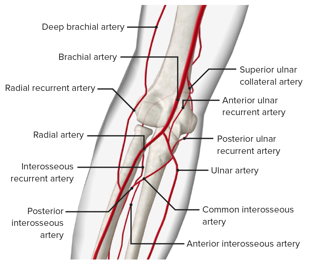
Blood Supply of Elbow joint
Nerve Innervation
The Nerve innervation of the elbow joint is slightly complicated and divided into many nerves in this order: the ulno-posterior part of the elbow is innervated by the ulnar nerve and some branches of medial antebrachial cutaneous nerve. The radial-posterior part of the elbow is innervated exclusively by the radial nerve. The ulno-anterior part of the elbow is innervated by the median nerve and the musculocutaneous nerve. [see fig 5]
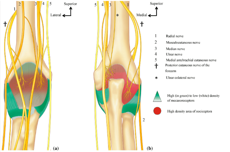
Nerve Innervation of the elbow joint
Capsule and Bursae
Like all synovial joints, The elbow joint is enclosed by a fibrous layer capsule, This joint capsule is weak anteriorly and posteriorly but is strengthened on each side by collateral ligaments, which stabilize the flexing and extending motion of the arm.
The synovial membrane lines the internal surface of the fibrous layer of the capsule and the intracapsular nonarticular parts of the humerus.
Also, there are many bursae in the elbow joint, but only a few have clinical importance, The three olecranon bursae are:
1- Intratendinous olecranon bursa: located within the tendon of the triceps brachii.
2- Subtendinous olecranon bursa: Which is located between the olecranon and the triceps tendon, reducing friction between the two structures during extension and flexion of the arm.
3- Subcutaneous olecranon bursa: Which is located between the olecranon and the overlying connective tissue (implicated in olecranon bursitis).
The bicipitoradial bursa (biceps bursa) separates the biceps tendon from, and reduces abrasion against, the anterior part of the radial tuberosity.
The synovial membrane lines the internal surface of the fibrous layer of the capsule and the intracapsular nonarticular parts of the humerus.
Also, there are many bursae in the elbow joint
Note
A bursa is a fluid-filled sac, that helps to reduce the friction between tissues of the body. Bursae are located near major joints such as shoulders, elbows, hips, and knees1- Intratendinous olecranon bursa: located within the tendon of the triceps brachii.
2- Subtendinous olecranon bursa: Which is located between the olecranon and the triceps tendon, reducing friction between the two structures during extension and flexion of the arm.
3- Subcutaneous olecranon bursa: Which is located between the olecranon and the overlying connective tissue (implicated in olecranon bursitis).
The bicipitoradial bursa (biceps bursa) separates the biceps tendon from, and reduces abrasion against, the anterior part of the radial tuberosity.
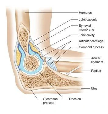
Elbow joint fibrous capsule & joint cavity
Ligaments
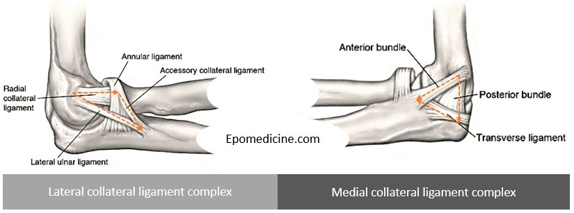
The Elbow joint has ligaments that connect the bones and contribute to its stability, which are:
Ulnar collateral ligament: It is triangular in shape, a ligament connecting the two bones involved in the articulation of the humeroulnar joint that extends from the medial epicondyle of the humerus to the coronoid process of the ulna. and is composed of three parts: an anterior, a posterior, and an inferior band.
Radial collateral ligament: a ligament connecting the two bones involved in the articulation of the humeroradial joint has a low attachment to the lateral epicondyle of the humerus. The distal fibers blend with the annular ligament that encloses the head of the radius.
Annular ligament: reinforces the joint by holding the radius and ulna together at their proximal articulation.
Quadrate ligament: helps to maintain constant tension during pronation and supination movements of the forearm. [we can see the ligaments in fig 6]
Bones and Ligament of Forearm
Relation
Anteriorly: brachialis, cubital fossa.
Posteriorly: anconeus, triceps.
Medially: ulnar n., common flexor origin.
Laterally: radial n., common extensor origin.
Posteriorly: anconeus, triceps.
Medially: ulnar n., common flexor origin.
Laterally: radial n., common extensor origin.
References
1) Moore K. L., Dalley, A. F., & Agur, A. M. R. (2014)- Clinically Oriented Anatomy 7th Edition (800-814 plates).
2) F. Netter: Atlas of Human Anatomy, 6th Edition, Elsevier Saunders (2014).
3) Elbow joint, iamh Gorman MSc, Kenhub.
https://www.kenhub.com/en/library/anatomy/elbow-joint
4) The Elbow Joint , Oliver Jones , Teachme Anatomy.
https://teachmeanatomy.info/upper-limb/joints/elbow-joint/
5) Physiotherapy Management of the Elbow, Physiopedia.
https://www.physio-pedia.com/Physiotherapy_Management_of_the_Elbow
References of images
Cover image: JOINT MOBILIZATION OF THE ELBOW JOINT COMPLEX - PHCN Online. https://i0.wp.com/phcn-online.com/wp-content/uploads/2021/05/image.jpeg?resize=615%2C400&ssl=1
fig 1: Bony anatomy of the joints of the elbow—anterior view. Musculoskeletal Key
https://musculoskeletalkey.com/wp-content/uploads/2016/08/B9781416058847000041_gr1.jpg
fig 2: Bony anatomy of the joints of the elbow—posterior view. Musculoskeletal Key
https://musculoskeletalkey.com/wp-content/uploads/2016/08/B9781416058847000041_gr2.jpg
fig 3: Anatomy of the elbow and movement of flexion-extension of the joint, ReasearchGate.
https://www.researchgate.net/publication/346430538/figure/fig1/AS:987100312772609@1612354462271/Anatomy-of-the-elbow-and-movement-of-flexion-extension-of-the-joint-33.ppm
fig 4: Elbow Joint: Anatomy [+video] - Lecturio Medical.
https://cdn.lecturio.com/assets/Arteries-of-the-elbow.png
fig 5: Schematic diagram of sensory innervation of the human elbow joint (A, anterior view; B posterior view). Articular ramifications originating from muscular branches were not included, Pierre Laumonerie.
https://www.researchgate.net/profile/Pierre-Laumonerie/publication/338210197/figure/fig1/AS:845406527315968@1578572031176/Schematic-diagram-of-sensory-innervation-of-the-human-elbow-joint-A-anterior-view-B.png
fig 6: Elbow ligaments: Simplified Anatomy | Epomedicine.
https://epomedicine.com/wp-content/uploads/2020/04/elbow-ligaments.jpg
fig 7: Joint: Elbow joint: Fibrous capsule & joint cavity | RANZCRPart1 Wiki | Fandom.
https://static.wikia.nocookie.net/ranzcrpart1/images/4/4d/Elbow_joint-1428E8201803B9EF10C.jpg/revision/latest?cb=20150326072420
