


Lumbrical Canal
By : Najah SabahDefinition
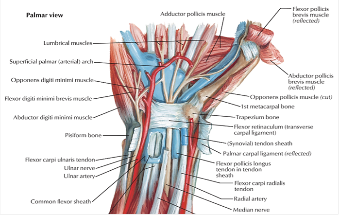
The lumbrical canal is a potential space surrounding the tendon of each lumbrical muscle and is normally filled with connective tissue. Proximally, it is continuous with one of the palmar spaces.(see figure 1)
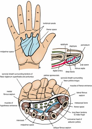
Its related structures :
Of course “ if we talk about the lumbrical canal . we must talk about the lumbrical muscles .
Definition
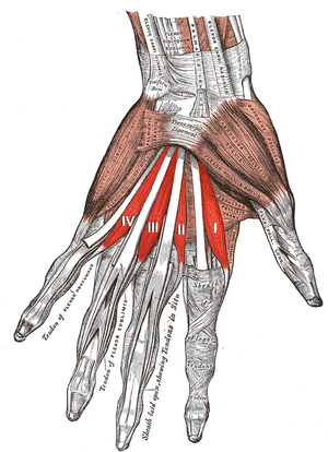
The lumbricals are deep muscles of the hand that flex the metacarpophalangeal joints and extend the interphalangeal joints. It has four, small, worm-like muscles on each hand. These muscles are unusual in that they do not attach to bone. Instead, they attach proximally to the tendons of flexor digitorum profundus and distally to the extensor expansions.[1]This muscle’s small cross-sectional area and measurements of the muscle’s strength in biomechanical studies suggest that this is a relatively weak muscle. This is especially true when the lumbrical is compared to the interosseous muscle, which has a similar function but is considerably stronger. The high number of muscle spindles in the lumbricals suggests that these muscles may have an important role in proprioceptive monitoring of the fingers. Furthermore, anatomical factors and the allocation of spindle fibers among the lumbricals suggests that this muscle is more involved in sensory feedback, which is important for precision pinch movements and precise manipulation of objects.
(see figure 2)
(see figure 2)
Structure
The lumbricals are four, small, worm-like muscles on each hand. These muscles are unusual in that they do not attach to bone. Instead, they attach proximally to the tendons of flexor digitorum profundus and distally to the extensor expansions.
Nerve
The first and second lumbricals (the most radial two) are innervated by the median nerve . The third and fourth lumbricals (most ulnar two) are innervated by the ulnar nerve .This is the usual innervation of the lumbricals (occurring in 60% of individuals). However 1:3 (median:ulnar - 20% of individuals) and 3:1 (median:ulnar - 20% of individuals) also exist. The lumbrical innervation always follows the innervation pattern of the associated muscle unit of flexor digitorum profundus (i.e. if the muscle units supplying the tendon to the middle finger are innervated by the median nerve, the second lumbrical will also be innervated by the median nerve). (see figure 3)
Artery
Four separate sources supply blood to these muscles: the superficial palmar arch, the common palmar digital artery, the deep palmar arch, and the dorsal digital artery.(see figure 4)
Function
The fact that the lumbrical muscles of the hand originate from tendons and insert into the extensor expansions, instead of bony structures, makes both of their attachment points quite mobile. That means the muscles are capable of two different actions. These are flexion at the metacarpophalangeal joints and extension in both the proximal and distal interphalangeal joints . The reason for the opposite actions is that the tendons cross the metacarpophalangeal joints on the palmar side but distally insert at the dorsal side of the finger. These combined movements play a role in complex movement of the hand (e.g. for holding a pen), and contribute to the general dexterity of the hand.
In addition, it is found that the lumbrical muscles of the hand contain many muscle spindles and have a large fiber length, which indicates that they likely play a role in proprioception.
(see video)
In addition, it is found that the lumbrical muscles of the hand contain many muscle spindles and have a large fiber length, which indicates that they likely play a role in proprioception.
(see video)
Clinical relevance
The lumbrical muscle is sometimes damaged in crush injuries to the hand. Following these injuries, adhesions may occur between the lumbrical muscle and the interossous muscle. Because the lumbrical passes volar to the inter-palmar plate ligament, whereas the interossous muscle passes dorsal, adhesions distal to the inter-palmar plate ligament limit the proximal movement of the interossous and lumbrical muscles. This deformity causes the patient to have intermetcarpal pain when making a fist. It can typically be treated by release of the involved muscles. The lumbrical muscles have also been shown to be relevant in carpal tunnel syndrome.
Lumbrical-plus finger : when the tendons of the flexor digitorum profundus detach distal to the origin points of the lumbricals an interesting phenomenon occurs: when trying to close the fist, the fingers strangely extend instead.
After detachment of the distal tendons, the lumbricals now serve as the new insertion surface of the flexor digitorum profundus. This means, even though the person consciously activates the flexor muscle, they actually move the lumbricals instead. And since the flexor digitorum profundus and the lumbricals are antagonists in the proximal interphalangeal and distal interphalangeal joints, the intended fist closure paradoxically leads to an extension of the fingers. This oddity is clinically referred to as the lumbrical-plus finger and can occur after injuries or amputations. (see figure 5)
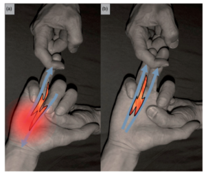
Stress test of lumbrical muscle tear. (a) Positive stress test: provocation of pain with passive extension of the ring finger while patient aims to close his fist. (b) No symptoms can be provoked when the middle finger is extended simultaneously, as the quadriga effect of the third lumbrical muscle disappears due to non-separation of the bipennate origins.
Treatment
Physiotherapy interventions include, first of all, restoring elasticity and then improving strength.
Reference:
1-LAWRENCE E. WINESKI, PhD, Snell’s Clinical Anatomy by Regions, TENTH EDITION,lumbrical canal , 298.
2- Lumbricals of the hand, physio-pedia, https://www.physio-pedia.com/Lumbricals_of_the_Hand.
2- Lumbricals of the hand, physio-pedia, https://www.physio-pedia.com/Lumbricals_of_the_Hand.
