Phalanges
By : Ruqayah AkramDefinition
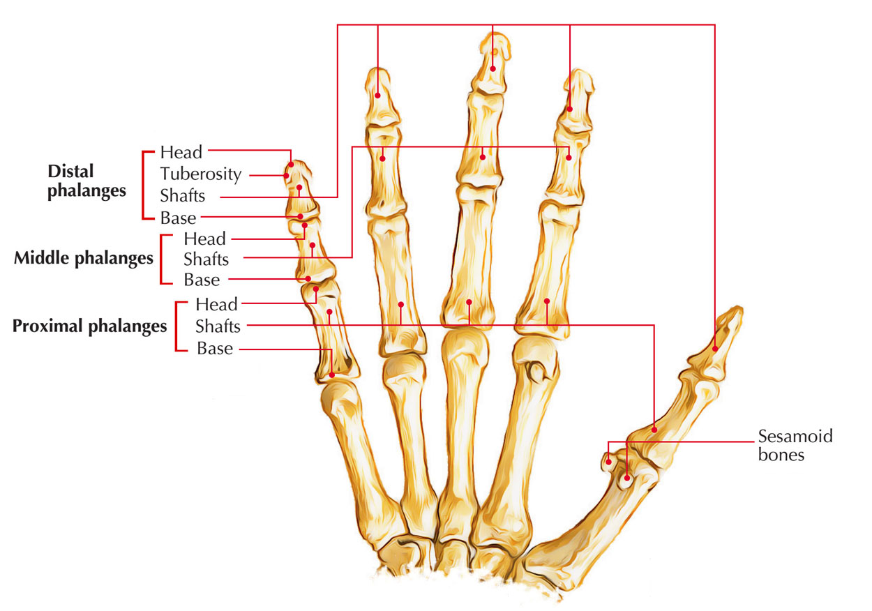
The phalanges are the bones of the digits of hand .
Type of bone
The phalanges are long bone .
Note : phalanges is plural for phalanx so the singular from is phalanx.
A)The thumb has two phalanges:
1.proximal phalanx
2.distal phalanx.
B)The rest of the digits have three phalanges:
1.Proximal phalanx
2.Middle phalanx
3.Distal phalanx
Note: The proximal phalanx is the largest one , compared to the distal and middle phalanges.
1.proximal phalanx
2.distal phalanx.
B)The rest of the digits have three phalanges:
1.Proximal phalanx
2.Middle phalanx
3.Distal phalanx
Note: The proximal phalanx is the largest one , compared to the distal and middle phalanges.
Parts of phalanges
Each phalanx has:
1.base (proximally)
2.shaft (body)
3.head (distally)
Notes:
1.The base of each proximal phalanx articulates with the head of the related metacarpal bone to form a metacarpophalangeal joint.
2.The head of each distal phalanx is non-articular and flattened into a crescent-shaped palmar tuberosity, which lies under the palmar pad at the end of the digit.
.jpg)
Blood Supply
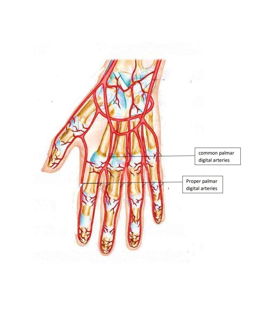
The blood supply of the phalanges is carried by the nutrient rami from palmar digital artery.
Note
rami: Latin term meaning branch
Clinical Note

clinical note
Fractures of phalanges
-Are common injuries.
-They occur more in males.
-The small finger is more affected compared to other fingers.
- Distal phalanges are common of the crushing injuries (e.g., when a finger is caught in a car door).
- These injuries are extremely painful because of the highly developed sensation in the fingers.
- A fracture of a distal phalanx is usually comminuted, and a painful hematoma (local collection of blood) soon develops.
- Fractures of the proximal and middle phalanges are usually the result of crushing or hyperextension injuries.
-Because of the close relationship of phalangeal fractures to the flexor tendons, the bone fragments must be carefully realigned to restore normal function of the fingers.
References
1-Keith L. Moore , Arthur F. Dalley A. M. R. Agur; Moore clinically oriented anatomy 7thedition;pp.673,679-681,683,687,771,773,789,812
2-Dr. Lawrence E. Wineski,PhD;SNELLIS CLINICAL ANATOMY BY REGIONS 10th edition;pp.39,215,237-239,315,363,422,429
3-Phalanges of the hand; Adrian Rad,BSc;Kenhub; https://www.kenhub.com/en/library/anatomy/the-phalanges
4-Phalanx Fractures of the Hand; Dalton J. McDaniel, Uzma H. Rehman.; National Library of Medicine; https://www.ncbi.nlm.nih.gov/books/NBK557625/
References of image
cover image: https://www.google.com/url?sa=i&url=https%3A%2F%2Fanatomy.app%2Fencyclopedia%2Fphalanges-of-hand&psig=AOvVaw2yEQIXfqkgBvW9vsiRPfp-&ust=1668250444538000&source=images&cd=vfe&ved=0CA8QjRxqFwoTCKic06r7pfsCFQAAAAAdAAAAABAJ
fig1:https://www.google.com/url?sa=i&url=https%3A%2F%2Fen.wikipedia.org%2Fwiki%2FPhalanx_bone&psig=AOvVaw2yEQIXfqkgBvW9vsiRPfp-&ust=1668250444538000&source=images&cd=vfe&ved=0CA8QjRxqFwoTCKic06r7pfsCFQAAAAAdAAAAABAO
fig2:https://www.google.com/url?sa=i&url=https%3A%2F%2Fboneandspine.com%2Fphalanx%2F&psig=AOvVaw0bUVgfqCgefzbY6sfOAXM6&ust=1668253413182000&source=images&cd=vfe&ved=0CA8QjRxqFwoTCLDforKGpvsCFQAAAAAdAAAAABAE
fig3:https://www.google.com/url?sa=i&url=https%3A%2F%2Fwww.illustratedverdict.com%2Ftemplate-arm-normal-anatomy&psig=AOvVaw0iotcx79eLTyeT0n1gMXiE&ust=1668253850724000&source=images&cd=vfe&ved=2ahUKEwjUj86CiKb7AhU6YPEDHVNcCC0QjRx6BAgAEAo
