Dorsal interossei manus muscles
By : Omar M. Subhi Altaie• NAME
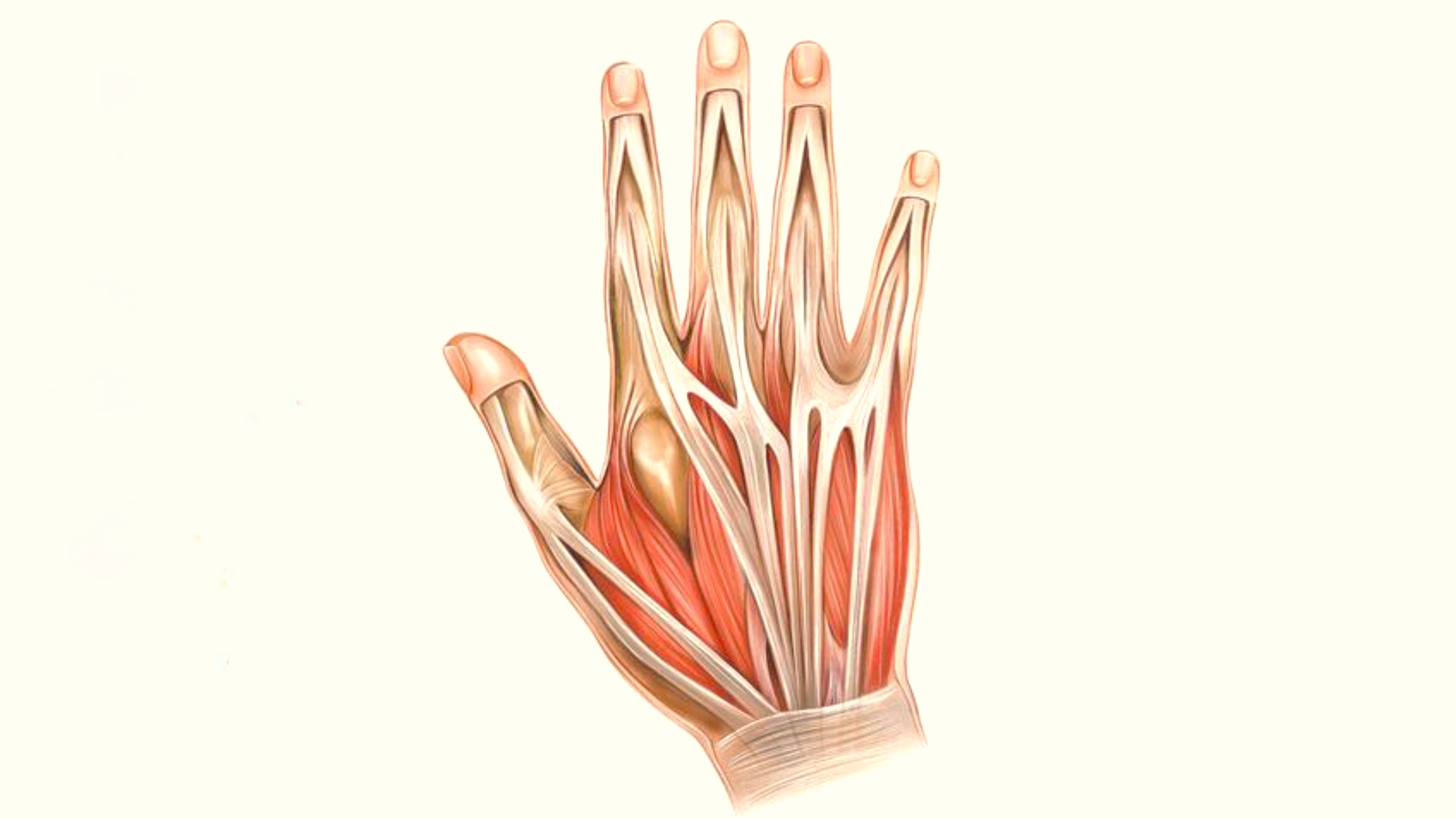
The name indicates that these four muscles are on the dorsal (back or posterior) of the hand (manus) .
The Latin word (interossei) means between the bones (ossei) of the metacarpals.
The Latin word (interossei) means between the bones (ossei) of the metacarpals.
• GENERAL
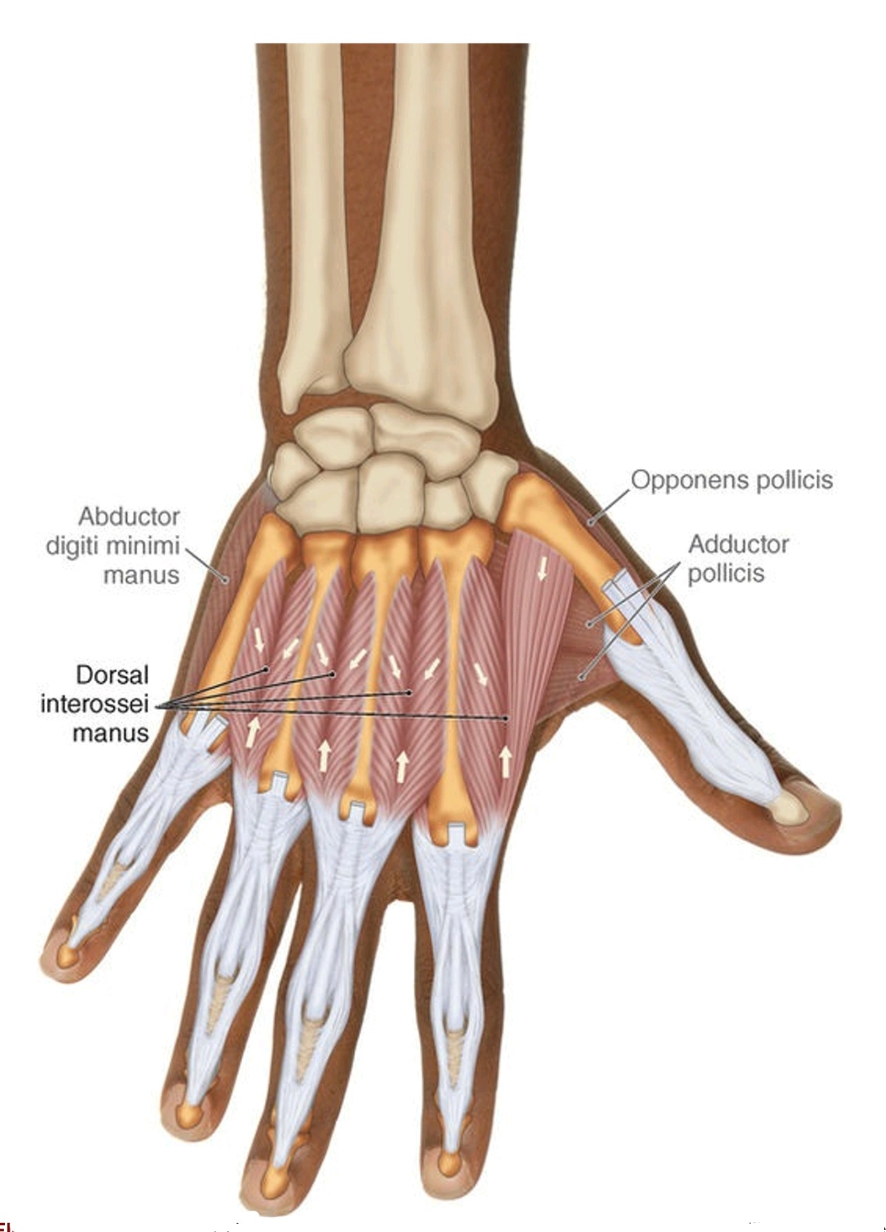
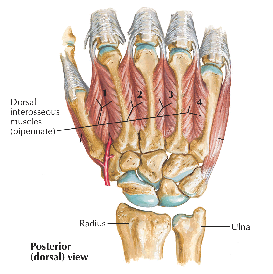
This muscle group is one of four muscle groups of the Central Compartment Group of the hand, they are four muscles named one, two, three, and four from lateral to medial ( radial to ulnar ) sides. they are bipennate in shape which means that the fibers are arranged just like a feather pattern with a tendon in the middle
They are deep to the palmar interossei (from palmer perspective) they are between the bones of metacarpals as the name indicates. Dorsal interossei muscles are on the dorsal surfaces of the metacarpal bones, in the interosseous compartment of the hand.
They apply their action at the metacarpophalangeal joints of the index, middle, and ring fingers, whereas the thumb and little fingers have their groups of muscles that are used exclusively on them.
They also cross the proximal and distal inter-phalangeal joints of the same fingers that they apply their actions on, they work with the other groups of the central compartment muscle groups to give the best function to the fingers.
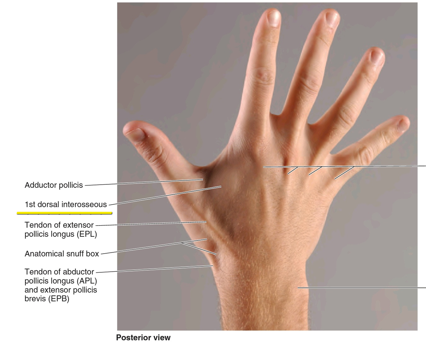
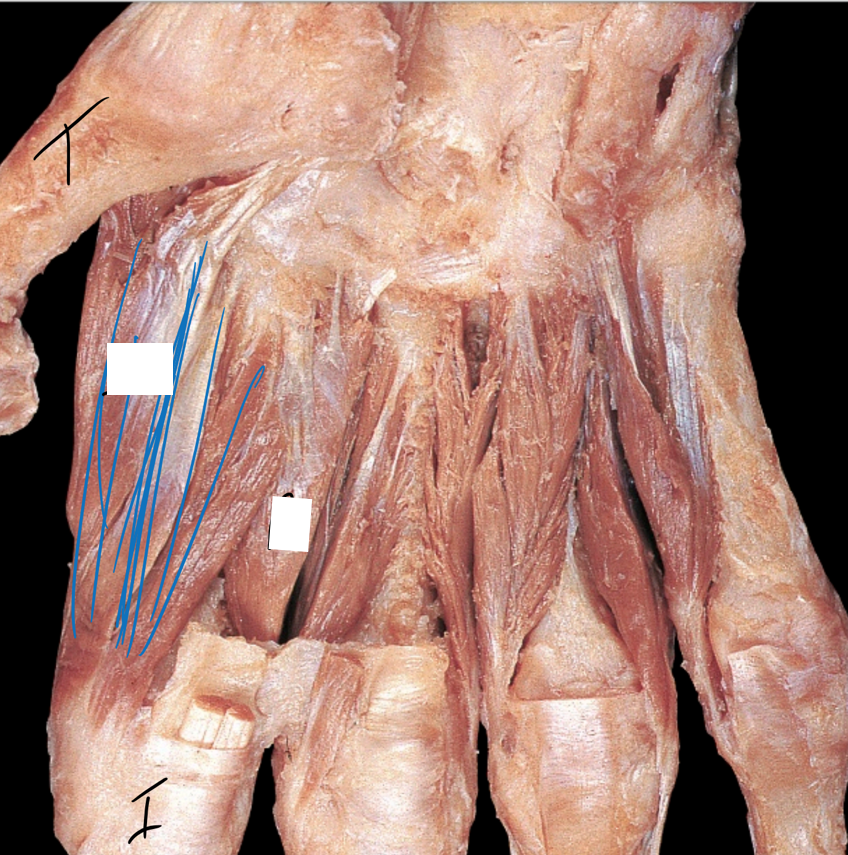
The first dorsal interosseous muscle can be easily palpated in the space somewhere between the thumb and index fingers. Whereas, it is also possible to palpate the other three interossei that consider being between the metacarpal bones and the tendons of the extensor digitorum muscle. (As shown in the figure below)
• SUPPLY
The nerve supply of the dorsal interossei manus is by the deep branchs of the Ulanr nerve (C8, T1) which is one of the terminal branchs of the brachial plexus.
The blood supply of the dorsal interossei manus is as follows:
• The first dorsal interosseus muscle is supplied by the first metacarpal branch that originates directly from the radial artery.
• Whereas the second, third, and fourth muscles are supplied from the second, third, and fourth dorsal metacarpal branches whiches originate from the dorsal carpal arch and run downward and reach these three muscles.
● ORIGIN AND INSERTION
◇ Origin
Dorsal interossei are bipennate muscles whiches feather-like patterns they can be seen in the dorsum aspect of the hand. The dorsal interossei consist of four muscles numbered from one to four, lateral to the medial side.
• The first dorsal interosseous muscle is the largest and strongest compared to other dorsal interossei muscles and is sometimes called the abductor indicis ( which is the index) as if it was an independent muscle. It arises from the medial shaft surfaces of the first lateral shaft surface of the second metacarpal bones.
• The second dorsal interosseous muscle originates from the medial and lateral surfaces of the 2nd and 3rd metacarpal bones respectively.
• The third dorsal interosseous muscle originates from the medial and lateral surfaces of the third and fourth metacarpal bones respectively.
• The fourth dorsal interosseous muscle originates from the medial and lateral surfaces of the fourth and fifth metacarpal bones respectively.
◇ Insertion
• The first dorsal interosseous muscle fibers travel obliquely and distally to continue as a tendon that inserts on the same lateral side of the base of the second proximal phalanx of the index, and its dorsal digital expansion.
• the second dorsal interosseous muscle fibers travel obliquely and distally into a tendon which inserts on the lateral side of the third digit on the base of its proximal phalanx and its dorsal digital expansion (hood).
• the tendon of the third dorsal interosseous muscle inserts on the medial side of the base of the same third digit,that the second interosseous inserts on, the proximal phalanx and its dorsal digital expansion (hood).
• Just like the third dorsal interosseous, the tendon of the fourth dorsal interosseous also inserts on the medial side of the fourth digit, by attaching to the base of its proximal phalanx and its dorsal digital expansion(hood).
• Movement and action
Their fibers' pattern faces away from the middle finger making its actions is pulling these metacarpal bones away from the midline and abducting these fingers, and that is the basci function of these muscles.
The first and second muscles cross the metacarpo-phalangeal joints of the index and middle fingers from the lateral side. They pull the proximal phalanx of these fingers laterally, away from the midline, and that is considered as abduction of the index finger.
But the second one abducts the middle finger to the outer side laterally. The third and fourth muscles cross the Metacarpophalangeal joints of the middle and ring fingers medially They pull the proximal phalanx of these fingers to the inner side, medially.
They all cross the Metacarpophalangeal joints of the fingers they are responsible for and abduct them at the same joints.
They can also flex the proximal phalanges of the index, middle, and ring fingers at the Metacarpoph-algeal joints of these fingers because their fibers anteriorly cross the joint.
The dorsal interossei can also extend the middle phalanx at its proximal interphalangeal joint and extend the distal phalanx at its distal interphalangeal joint, the reason behind this is because its fibers travel distally to cross these joints of the fingers two, four, and five respectively from the posterior side of them.
• Reverse actions
As we know the normal action of any muscle is when the proximal attachment of the origin of that muscle is fixed and the distal attachment which is the insertion is the movable portion, but when the opposite of that happens we call it Reverse action
When the index finger is fixed,(and that might be because the person is holding something in his hand) the head of the first dorsal interosseus manus that attaches to the thumb pulls the thumb medially from where it is attached and posteriorly toward the index finger, causing flexion and adduction of the thumb those actions occur at the carpometacarpal joint.
Extending the proximal phalanges at the interphalangeal joints refers to extending the proximal phalanx at the Proximal interphalangeal joint and extension of the middle phalanx occurs at the distal interphalangeal joint.
• ANTAGONIST FUNCTIONS
The dorsal interossei slow and control adduction and extension of fingers two through five at the Metacarpophalgeal joints and stabilize the joints.
They also slow flexion of the phalanges of the fingers two, four, and five at the proximal and distal interphalangeal joints. They stabilize these joints and the Carpometacarpal joint of the thumb.
They also slow flexion of the phalanges of the fingers two, four, and five at the proximal and distal interphalangeal joints. They stabilize these joints and the Carpometacarpal joint of the thumb.
• Examination
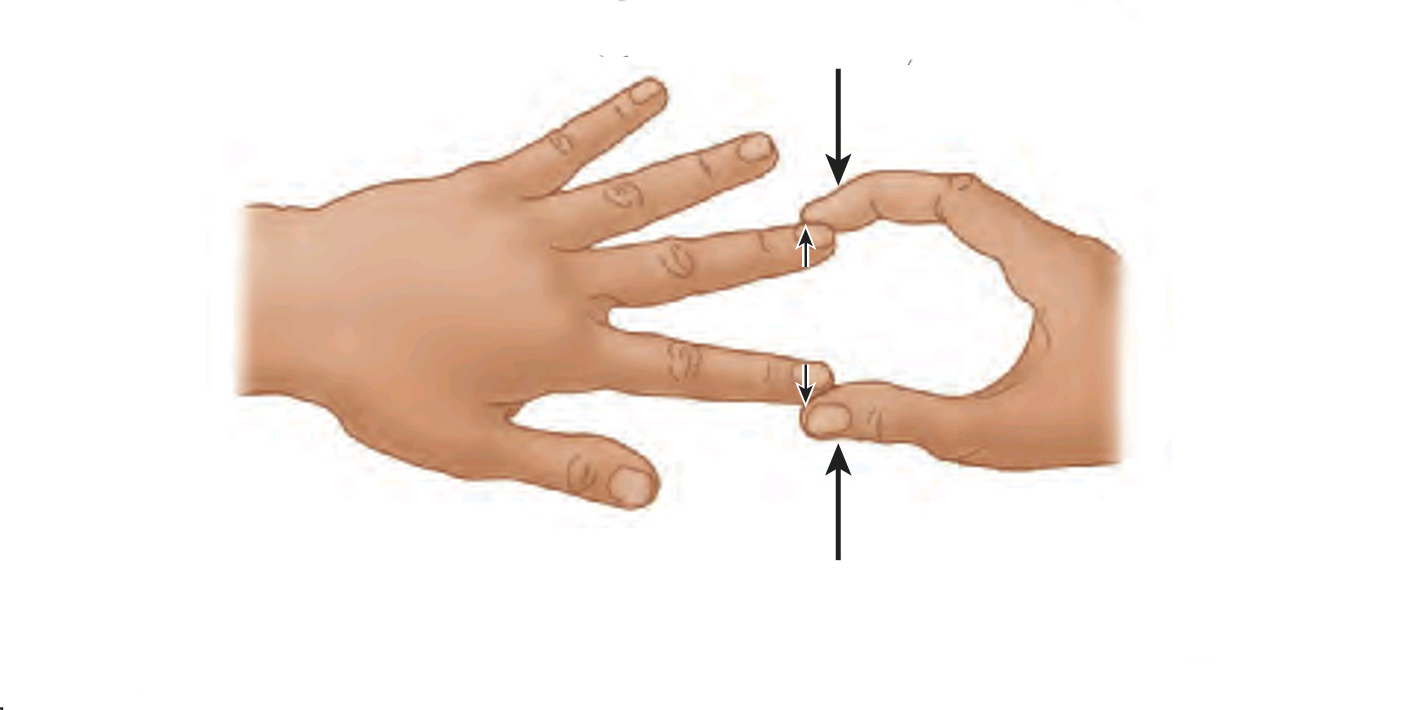
We can test these muscles by asking the patient to abduct (spread) his two adjacent fingers against the resistance of the examiner who is preventing him from doing that. If he can do that easily, his dorsal interossei are intact .
• Clinical notes
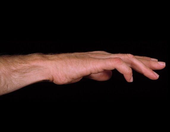
As we know the medial two of these muscles are innervated by the deep branch of the Ulnar nerve , and injury occurs that never will cause weekness and paralysis to these two muscles; thus the patient will not be able to abduct his fingers.
Furthermore, after that injury, a deformity called ulner claw hand deformity when the affected fingers are flexed at the interphalangeal joints and extended at the Carpometacarpal joints.
That deformity is caused by a weakness in the third and fourth lumbricals and the interossei manus. The other two fingers, the index, and middle fingers are not very affected by the ulnar claw deformity; because their two lumbricals innervated by the median nerve which is intact in this case.
• RELATIONS
The dorsal interossei muscles can be seen in the dorsum of the hand. From an anterior perspective they lie deep to the palmar interossei, filling the space between the metacarpal bones.
The first one lies deep to the adductor pollicis at the lateral side of the hand.
the extensor digitorum tendons passing over the dorsal interossei from the posterior aspect.
The dorsal interossei manus are superficial and located between the bones.
The first one lies deep to the adductor pollicis at the lateral side of the hand.
the extensor digitorum tendons passing over the dorsal interossei from the posterior aspect.
The dorsal interossei manus are superficial and located between the bones.
● REFERENCES
• JOSEPH E. MUSCOLINO THE MUSCULAR SYSTEM MANUAL The Skeletal Muscles of the Human Body FOURTH EDITION 420-421, 435-439
• Keith L. Moore, Arthur F. Dalley, Anne M. R Clinically Oriented Anatomy (7th Edition) page779
• Snell's clinical anatomy by Region's (10th edition) page 321
• Dorsal interossei of the hand Physiopedia https://www.physio-pedia.com/Dorsal_Interossei_of_the_Hand
• Anatomy, Shoulder and Upper Limb, Hand Dorsal Interossei Muscle national library of medicine, Valenzuela M, Bordoni B. https://www.ncbi.nlm.nih.gov/books/NBK536922/#:~:text=The%20dorsal%20interosseous%20muscles%20are,extension%20of%20the%20interphalangeal%20joints.
● IMAGES REFERENCES
• Cover image by https://www.alamy.com/illustration-of-the-muscles-of-the-hand-this-is-a-dorsal-superficial-view-of-the-muscles-of-the-hand-illustration-from-asklepios-atlas-of-the-human-image334945452.html
• Fig1 JOSEPH E. MUSCOLINO THE MUSCULAR SYSTEM MANUAL The Skeletal Muscles of the Human Body FOURTH EDITION page 436
• Fig3 Frank H. Netter, Atlas of Human Anatomy (7th edition) plate 455
• Fig2 & fig5 Testing interossei (ulnar nerve). Dorsal interossei muscles FIGURE 6.80, Keith L. Moore, Arthur F. Dalley, Anne M. R Clinically Oriented Anatomy (7th Edition) page 779, 789
• Fig4 by nick_rodriguez75 https://quizlet.com/269454401/interossei-cadaver-diagram/
• Fig6 from https://www.chiropractic-in-malaysia.com/blog/ulnar-nerve-palsy-claw-hand
