


Lumbrical Manus Muscles
By : Omar M. Subhi Altaie• NAME
The first part of the name indicates the worm-like shape of the muscles and the Latin word " manus " means the hand.
• GENERAL
Four worm-like muscles that are present in the Central Compartment Group of the hand, cross the metacarpophalangeal joints and flex the fingers at them. They cross and extend the fingers at the proximal and distal interphalangeal joints of the lateral four fingers of the hand .they attach to the tendons of the extensor digitorum muscle whiches reaches the distal phalanges of the fingers.

• SUPPLY
The nerve supply of the lumbricals manus muscles is as follows :
1. The first two lumbricals from the lateral side are innervated by the lateral palmar digital branches of the median nerve
2. The third and fourth lumbricals innervated by a deep branch of the Ulnar nerve
And here is a great way to memories that by a mnemonic :
• 1 st and 2 nd lumbricals U lnar ( one to You )
• 3rd and 4th lumbricals M edian (three for Me )
The blood supply of the lumbricals is by branches from arches whiches originate from the ending of Radial and Ulnar arteries or branches from the arteries themself; they are several separated sources :
1. The first and second dorsal metacarpal arteries ( branches from the dorsal carpal arch) and dorsal digital arteries, supply the
first and second lumbricals muscles.
2. The third and fourth dorsal digital arteries (branches from the dorsal carpal arch), as well as the second and third common palmar digital arteries (which are branches of the superficial palmar arch) supply the third and fourth lumbricals muscles.
• ORIGIN AND INSERTION
◇ ORIGIN
The lumbrical muscles of the hand are named one to four from the lateral to the medial. Each lumbrical muscle has one or two sites of origin from adjacent tendons of the flexor digitorum profundus muscle that presents in the forearm but its long tendons reach the distal phalanges of the fingers to apply their actions on them.
The first two Lumbricals are as noticed unipennate, and that means their fibers do not the feather-like pattern so they arise from one side of the origin- tendons otherwise other two medial lumbricals are bipennate; which is having the feather pattern and arising from two sides of origin-tendons.
○ The first lumbrical (unipennate) from the lateral side and palmar surface of the tendon of the flexor digitorum profundus muscle at the beginning of the hand superficial to the carpal bones, and that tendon is in its way inserted in the distal phalanx of the second metacarpal bone which is the index finger.
○ The second lumbrical (unipennate) from the lateral side and palmar surface of the tendon of the flexor digitorum profundus muscle at the beginning of the hand superficial to the carpal bones, and that tendon is in its way inserted in the distal phalanx of the third metacarpal bone which is the middle finger.
○ The third lumbrical (pennate) so arises from the lateral and medial sides of the tendon of the flexor digitorum profundus muscle at the beginning of the hand superficial to the carpal bones, and that tendon is in its way inserted into the distal phalanx of the fourth metacarpal bone which is the ring finger.
○ The fourth lumbrical (pennate) so arises from the lateral and medial sides of the tendon of the flexor digitorum profundus muscle at the beginning of the hand superficial to the carpal bones, and that tendon is on its way inserted into the distal phalanx of the fifth metacarpal bone which is the little finger.

◇ INSERTION
Each of the lumbrical muscles passes toward the end of the hand in the distal phalanges, along the lateral side of the finger that its responsible for, anterior to the deep transverse metacarpal ligament. The lumbricals then extend obliquely (as the pattern of the direction of its fibers) to attach to the lateral sides of the extensor expansion of the Extensor digitorum muscle of the digit that muscle is meant to control
Each of the lumbrical muscles passes toward the end of the hand in the distal phalanges, along the lateral side of the finger that its responsible for, anterior to the deep transverse metacarpal ligament. The lumbricals then extend obliquely (as the pattern of the direction of its fibers) to attach to the lateral sides of the extensor expansion
Note
(some small triangular aponeurosis that covers the dorsum aspect of the proximal phalanx of the finger to connect between two tendons, it is near to its base with the metacarpophalangeal joints) 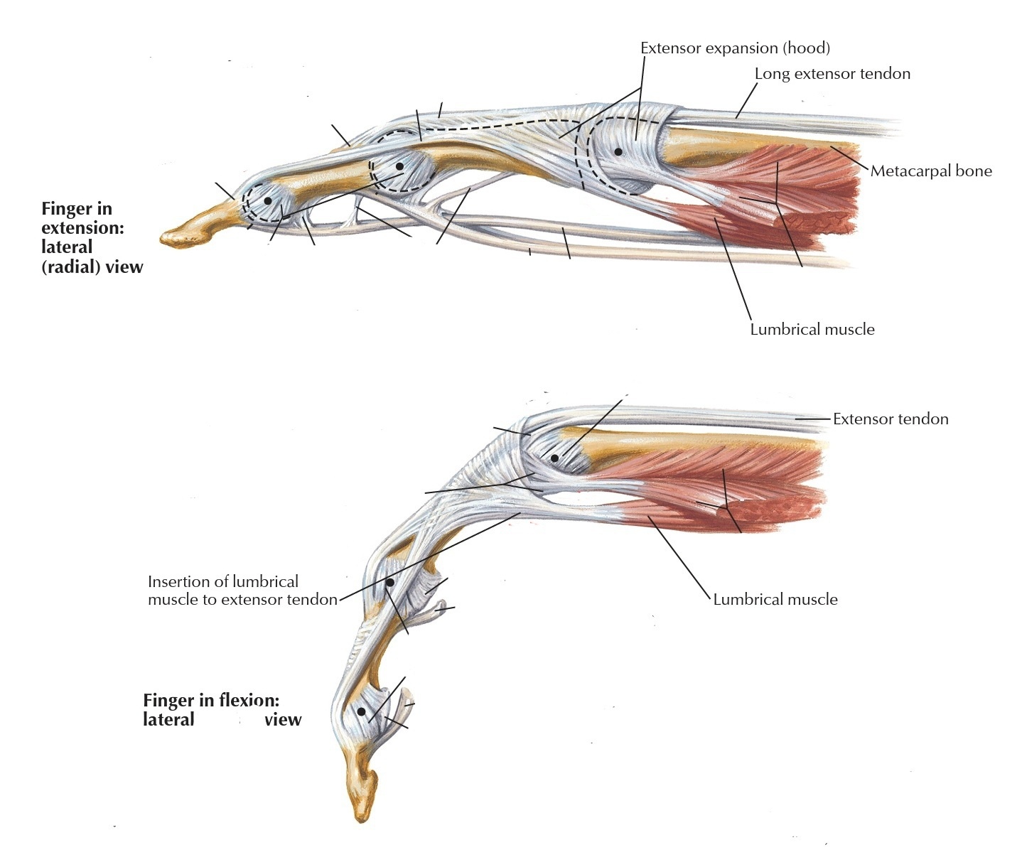
• MOVEMENT & ACTION
The lumbricals manus muscles attach to the tendons of the extensor digitorum muscle in the first quarter of the proximal phalanx of the fingers. When it contracts it pulls the distal end of the tendons which attach to the distal phalanx, of the fingers.
They posteriorly cross the proximal and distal interphalangeal joints and thus when it contracts, they extends the middle and distal phalanges at proximal and distal interphalangeal joints respectively.
From the anterior aspect, they cross the metacarpophalangeal joints ; so if these fibers contract, they could flex the proximal phalanx at those joints.
From the lateral side they cross the metacarpophalangeal joints of the fingers, so they have the ability to move these fingers in the direction depending on the side that they crossing the fingers; the first two Lumbricals cross the index and middle fingers' metacarpophalangeal joints laterally, thus if these muscles contract, they will move them away from the midline and that is ABduction of these fingers,but the third and fourth lumbricals move the ring and little fingers laterally which is now toward the midline and that considered as adduction of these fingers.
• REVERSE ACTIONS
These muscles can extend the proximal and middle phalanges at the proximal and distal interphalangeal joints .
The reverse action of a finger at the Metacarpophalangeal joint occurs when the metacarpal moves slightly to the proximal phalanx of the finger at the same joint, and the distal attachment should be fixed for some reason, like when a person is holding some weights in his hands.
The reverse action of a finger at the Metacarpophalangeal joint occurs when the metacarpal moves slightly to the proximal phalanx of the finger at the same joint, and the distal attachment should be fixed for some reason, like when a person is holding some weights in his hands.
• ANTAGONIST FUNCTIONS
slows flexion of fingers two through five at the interphalangeal joints against the muscles that flex them, and stabilize these joints.
slows and controls the extension, adduction, and abduction of fingers two through five at the Metacarpophalgeal joints and stabilizes these joints.
slows and controls the extension, adduction, and abduction of fingers two through five at the Metacarpophalgeal joints and stabilizes these joints.
• Examination
To test the lumbrical muscles, with the hand in supine position, ask the patient to flex the metacarpophalangeal joints while keeping the interphalangeal joints extended. The examiner uses his finger to apply some resistance along the palmar surface of the proximal phalanx of each digits two through five individually.
to test the extension of the interphalangeal joints resistance may also be applied separately on the dorsal surface of the middle and distal phalanges of these fingers, also while flexion of the Metacarpophalangeal joints is maintained.
to test the extension of the interphalangeal joints resistance may also be applied separately on the dorsal surface of the middle and distal phalanges of these fingers, also while flexion of the Metacarpophalangeal joints is maintained.
• CLINICAL NOTES
○ Lower Lesions of Brachial Plexus (Klumpke’s Pals)
Lower lesions of the brachial plexus are usually followed by injuries caused by excessive abduction of the arm, as we can see that when someone is falling from a high building for example, he tries to save himself by attaching to any near object, so because of the very-fast falling, he cannot control the movement of his arms and might lead him to this injury.
the ulnar and median nerves originate in the lower of the brachial plexus to supply all the lumbricals muscles of the hand. Because the person cannot control his lumbricals he will have a clawed appearance caused by hyperextension at the metacarpophalangeal joints and flexion of the interphalangeal joints. The extensor digitorum is unopposed by the lumbricals and interossei are not used to extend the metacarpophalangeal joints and opposite the actions and the flexor digitorum superficialis and profundus are unopposed by the lumbricals and interossei thus the middle and distal phalanges, will be in the flexion position.
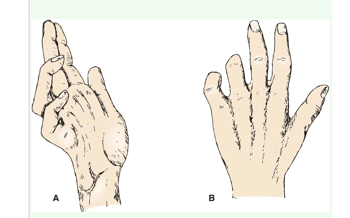
○ Median nerve palsy
Because the first two lumbricals are innervated by the median nerve so in this case the median nerve is injured no flexion is possible at the interphalangeal joints of the index and middle fingers, even though a weak flexion of the metacarpophalangeal joints of these fingers can be occurred by the interossei.
When the doctor asks the patient to make a fist, the index cannot be flexed, and the middle finger is half flexed, whereas the ring and little fingers flex, (because of the ulnar innervation). However, the latter two fingers are weakened by the loss of the flexor digitorum superficialis because its tendons reach these fingers and apply their actions to them.
the hand looks flattened and similar to that of a great ape, with the deformity most seen on the lateral side aspect of the hand. Thus, this posture of the upper limb resulting from a median nerve lesion is often referred to as “ape hand” as seen in the figure below.
Lower lesions of the brachial plexus are usually followed by injuries caused by excessive abduction of the arm, as we can see that when someone is falling from a high building for example, he tries to save himself by attaching to any near object, so because of the very-fast falling, he cannot control the movement of his arms and might lead him to this injury.
the ulnar and median nerves originate in the lower of the brachial plexus to supply all the lumbricals muscles of the hand. Because the person cannot control his lumbricals he will have a clawed appearance caused by hyperextension at the metacarpophalangeal joints and flexion of the interphalangeal joints. The extensor digitorum is unopposed by the lumbricals and interossei are not used to extend the metacarpophalangeal joints and opposite the actions and the flexor digitorum superficialis and profundus are unopposed by the lumbricals and interossei thus the middle and distal phalanges, will be in the flexion position.
○ Median nerve palsy
Because the first two lumbricals are innervated by the median nerve so in this case the median nerve is injured no flexion is possible at the interphalangeal joints of the index and middle fingers, even though a weak flexion of the metacarpophalangeal joints of these fingers can be occurred by the interossei.
When the doctor asks the patient to make a fist, the index cannot be flexed, and the middle finger is half flexed, whereas the ring and little fingers flex, (because of the ulnar innervation). However, the latter two fingers are weakened by the loss of the flexor digitorum superficialis because its tendons reach these fingers and apply their actions to them.
the hand looks flattened and similar to that of a great ape, with the deformity most seen on the lateral side aspect of the hand. Thus, this posture of the upper limb resulting from a median nerve lesion is often referred to as “ape hand” as seen in the figure below.
○ Ulnar nerve palsy
The third and fourth lumbricals innervated by the ulnar nerve, so when the nerve is injured they cannot be functioning to apply their action on the fingers, which can be seen when examining the patient by asking him to make a fist or gripping a paper between these fingers; adduction or flexion cannot be occurred by these muscles whiches lead to hyperextended metacarpophalangeal joints .
The interphalangeal joints are flexed, because of again the paralysis of these two muscles and interosseous muscles, which are normally meant to be extending these joints.
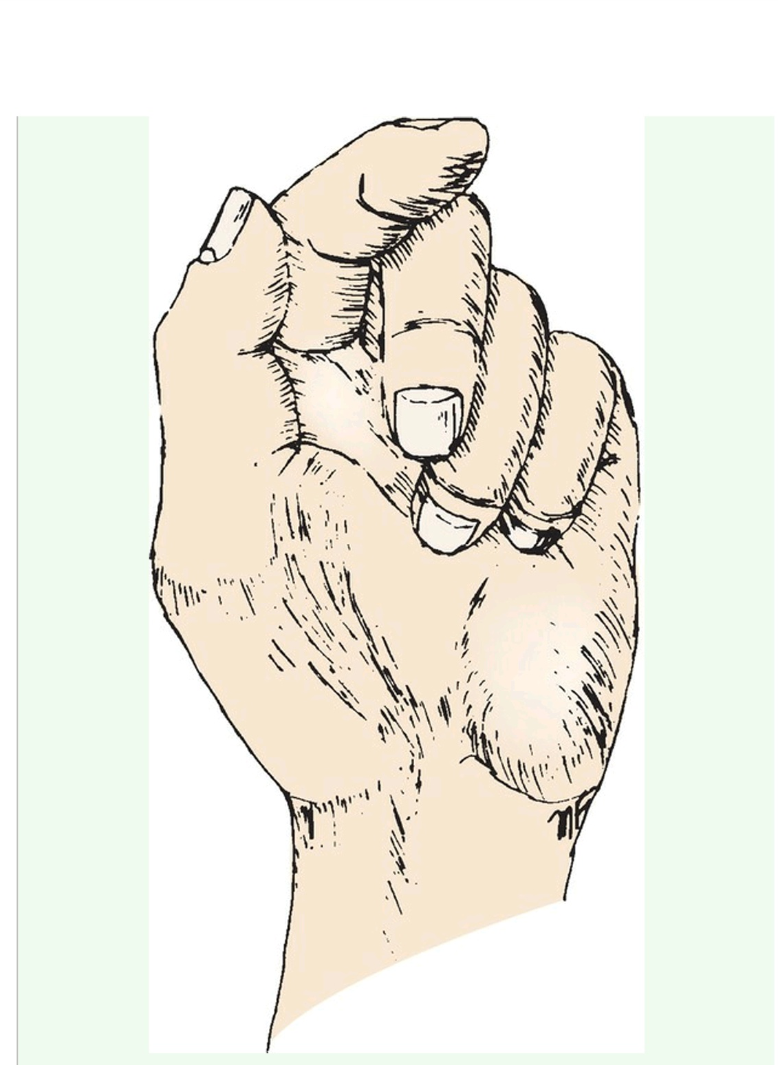
○ The "LUMBRICAL PLUS" finger
This "lumbrical plus" phenomenon occurs most commonly in the middle finger or index. When the origin of the lumbricals is detachment, so when the person is trying to flex his fingers they extend instead. The surface of the tendons of flexor digitorum profundus is now the insertion of these muscles.
When the person with this case trying to flex the index finger, he actually can easily control and prevent the pull against the flexion through the lumbrical by not contracting flexor digitorum profundus when he flexes the proximal interphalangeal joint with flexor digitorum
Superficialis that its tendon is reached.
But with the person trying to flex one of the three medial fingers he cannot fully control the pulling on flexor digitorum profundus to the affected finger when he fully flexes the normal fingers into the palm when gripping: that because he cannot control the specific tendon of that muscle to contract by independently, the tendons to these fingers came from the same muscle belly in the forearm, whenever it contracts, it contracts at once.
A lumbrical-plus finger results from the abnormality of biomechanics and only surgery to reconnect the profundus can resolve this paradoxical of the IP joints.

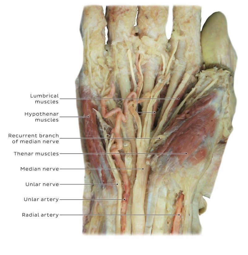
• RELATIONS
From the palmar side not very deep, under the palmar fascia. They get to the deeper layer when approaching the phalanges of the fingers, then they get posterior of these phalanges and attach to the tendons of the extensor digitorum muscle.
Deep to the first and second lumbricals is the adductor pollicis muscle, but deep to the third and fourth ones are the metacarpals bones and palmar interossei muscles between them.
Deep to the first and second lumbricals is the adductor pollicis muscle, but deep to the third and fourth ones are the metacarpals bones and palmar interossei muscles between them.
● REFERENCES
• JOSEPH E. MUSCOLINO THE MUSCULAR SYSTEM MANUAL The Skeletal Muscles of the Human Body FOURTH EDITION 420-421, 427-430
• Keith L. Moore, Arthur F. Dalley, Anne M. R Clinically Oriented Anatomy (7th Edition) page 779-792
• Snell ANATOMY BY REGIONS 10th Edition 320, 358-363
• Chihiro Yokochi, E. Lutejen-Drecoll, and Johannes W. Rohen Color Atlas of Human Anatomy 7th Edition page
• Lumbricals of the Hand https://www.physio-pedia.com/Lumbricals_of_the_Hand#:~:text=The%20lumbricals%20are%20deep%20muscles,do%20not%20attach%20to%20bone.
• Lumbrical muscles of the hand Author: Gordana Sendić MD • Reviewer: Egle Pirie Last reviewed: July 27, 2022 https://www.kenhub.com/en/library/anatomy/lumbrical-muscles-of-the-hand
• THE "LUMBRICAL PLUS" FINGER ATHOL PARKES, GLASGOW https://online.boneandjoint.org.uk/doi/pdf/10.1302/0301-620X.53B2.236
● IMAGES REFERENCES
• cover image by https://www.alamy.com/illustration-of-the-muscles-of-the-hand-this-is-a-dorsal-superficial-view-of-the-muscles-of-the-hand-illustration-from-asklepios-atlas-of-the-human-image334945461.html
• Fig1 JOSEPH E. MUSCOLINO THE MUSCULAR SYSTEM MANUAL The Skeletal Muscles of the Human Body FOURTH EDITION FIGURE 9-15 Anterior view of the right lumbricals manus. The adductor pollicis has been ghosted in. page 428
• Fig2 & fig3 Frank H. Netter, Atlas of Human Anatomy (7th edition) plate 451, 454
• Fig4 Radial artery traversing the anatomical snuffbox. Radial artery is the most lateral arterial vessel you will see in the wrist. Radial artery Author: Natalie Joe • Reviewer: Jerome Goffin Last reviewed: June 07, 2022, https://www.kenhub.com/en/library/anatomy/radial-artery
• Fig5 & fig6 Snell ANATOMY BY REGIONS 10th Edition Figure 3.62 Ulnar nerve palsy. A. Anterior view. B. Posterior view. page ,363 Figure 3.61 Median nerve palsy, anterior view page 358
• Fig6 The lumbrical fingers THE "LUMBRICAL PLUS" FINGER ATHOL PARKES, GLASGOW https://online.boneandjoint.org.uk/doi/pdf/10.1302/0301-620X.53B2.236 Figure 3 and 4
