


Adductor Pollicis Muscle
By : Omar M. Subhi Altaie• NAME
The first part of the name indicates that the main action of this muscle is adduction (moves something toward the midline) " adductor ", and the second part indicates that it applies that action to the thumb finger "pollicis".
• GENERAL
A fan-like shaped muscle that presents laterally in the adductor component of the hand.
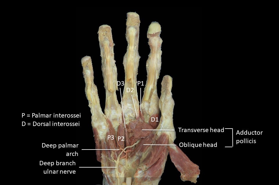
It is composed of two heads Oblique and transverse heads that differ in the orientation of their fibers, the radial artery passing in between the two just before it enters the palm.
It is analogous to the adductor hallucis that is present in the foot.
There is a lot of variation regarding the region of the oblique head of the adductor pollicis. The oblique head often has a fasciculus (which is a group of muscle fibers) that joins with the flexor pollicis brevis and is called the deep head of the flexor pollicis brevis.
One of two sesamoid bones that are present in the thumb is in the oblique head of the adductor pollicis muscle and it is the medial one of them.
This muscle represents the strong tissue of the thumb, in addition to the first dorsal interosseus manus muscle.
The adductor pollicis attached to the dorsal digital expansion of the thumb is small and often absent. If it is absent, the adductor pollicis will not have the ability to extend the distal phalanx of the thumb at the interphalangeal joint of the thumb.
It also crosses the saddle Carpometacarpal joint of the thumb and the interphalangeal joint as well.
It is composed of two heads Oblique and transverse heads that differ in the orientation of their fibers, the radial artery passing in between the two just before it enters the palm.
It is analogous to the adductor hallucis that is present in the foot.
There is a lot of variation regarding the region of the oblique head of the adductor pollicis. The oblique head often has a fasciculus (which is a group of muscle fibers) that joins with the flexor pollicis brevis and is called the deep head of the flexor pollicis brevis.
One of two sesamoid bones that are present in the thumb is in the oblique head of the adductor pollicis muscle and it is the medial one of them.
This muscle represents the strong tissue of the thumb, in addition to the first dorsal interosseus manus muscle.
The adductor pollicis attached to the dorsal digital expansion
Note
which is aponeurosis tissue that covers the phalanges of the fingers and their base with a metacarpophalangeal jointIt also crosses the saddle Carpometacarpal joint of the thumb and the interphalangeal joint as well.

• SUPPLY
The nerve supply of the adductor pollicis muscle is by the Deep branch of the ulnar nerve(C8, T1) after passing the hypothenar muscle group on the medial side of the hand and ends in the adductor pollicis laterally.
The blood supply of the adductor pollicis muscle is by branches of the Radial Artery
The blood supply of the adductor pollicis muscle is by branches of the Radial Artery
Note
which is one of two terminal branches of the Brachial Artery in the cubital fossa• ORIGIN AND INSERTION
The two heads of this muscle originate from different sites to make its triangular shape.
◇ Origin
Transverse head: originates mostly from the shaft of the second metacarpal bone which continues to become the index finger.
Oblique head: originates from the capitate bone (the second medial bone of the second row of carpal bones) and the base of the second and third metacarpal bones.
◇ Insertion
The two heads get closer to each other and insert as the fibers travel laterally, forming the tendon which often contains a sesamoid bone. By that tendon, it inserts on the base of the medial and anterior aspect of the proximal phalanx of the thumb as well as the extensor expansion (hood).
• MOVEMENT & ACTION
The adductor pollicis crosses the three joints that the thumb contains to them. The first one is the carpometacarpal joint (between the carpal bones and the metacarpal bone of the thumb) it crosses the joint posteriorly by attaching from the posteriorly and slightly medial side on the palm and distally attached anterolaterally onto the thumb.
When the thumb is in an abducted position the adductor pollicis contracts, it pulls the metacarpal bone of the thumb bringing it back to be at zero degrees with the palmar surface. This action is called the adduction of the thumb.
The adductor pollicis also crosses the first Carpometacarpal joint of the thumb from medially on the hand to attach laterally onto the outer surface of the metacarpal of the thumb. When it contracts, it Flexes the thumb and that can be seen when the metacarpal bone of the thumb is pulled medially to the anterior to the palm flexing the Carpometacarpal joint of the thumb , where toward its fixed attachment (origin).
It also crosses the first Metacarpophalangeal joint of the thumb medially. So when it contracts the adductor pollicis also flexes the proximal phalanx of the thumb at the first at the same joint.
it reaches with its distal attachment the dorsal digital expansion , and that makes it crosses the Interphalangeal joint of the thumb on the lateral side, so, in the flexion position, the adductor pollicis can be involved in the extension of the thumb by extending the distal phalanx at the interphalangeal joint of the thumb.
Additionally, when know adduction is one of the components of the opposition of the thumb, thus the adductor pollicis is involved in the opposition movement that occurs at the Carpometacarpal joint particularly.
That action can be used when gripping something between the thumb and that finger that the opposition is done with.
• REVERSE ACTION
As we know the normal action of any muscle is when the proximal attachment of the origin of that muscle is fixed and the distal attachment which is the insertion is the movable portion, but when the opposite of that happens we call it Reverse action
The reverse action occurs when the (trapezium or the origin place) moves toward the metacarpal bone of the thumb at the Carpometacarpal joint. That is because the metacarpal bone is fixed, for some particular reason. Movement of the trapezium could make the entire carpus (carpal bones) and some near places of the forearm so move too.
The other reverse action of the thumb occurs at the metacarpophalangeal joint when the fixed part here is the proximal phalanx and the metacarpal bone moves relative to it the thumb at that joint.
The last reverse action occurs at the interphalangeal joint when the distal phalanx is fixed and the proximal one is moving towards it in the plane that makes the extension action.
• ANTAGONIST FUNCTION
Like with any other muscle it stabilizes the joints that it crosses and they are as mentioned: Carpometacarpal , Metacarpophalangeal , and interphalangeal joints.
The adductor pollicis slows abduction of the CMC joint of the thumb as it is considered the opposite of adduction action.
It also slows the extension of the CMC and MCP joints of the thumb due to the flexion that this muscle is capable of doing.
It slows and controls the flexion of the IP joint of the thumb because of the extensor that occurs at this point, which this muscle is responsible for.
• EXAMINATION
By placing a sheet of paper between the thumb and the index fingers and asking the patient to grip and make a strong pinch, the doctor now tries to pull that paper back to him, if the patient fails to grip the paper strongly he might be diagnosed with Froment’s Sign
• CLINICAL NOTES
Froment’s Sign
Froment's sign is a physical examination that can be applied to the hand for testing the blockage and palsy of the ulnar nerve, and that can cause functionless and muscle weakness of the pinch grip that is done by the patient.
Froment's sign is followed by damage to the ulnar nerve due to an accident or injury to the nerve, which innervates the muscle.
The adductor pollicis can adduct the thumb as well as the extent of the only interphalangeal joint of the thumb. The flexor pollicis longus from the forearm (which receives its innervation by the median nerve), will substitute for the adductor pollicis and cause the thumb to get into hyperflexion.
palsy of the Ulnar nerve can be seen after dysfunction at the cervical spine, elbow cubital tunnel syndrome (compression of the nerve behind the medial epicondyle near the elbow joint), or at the wrist (Guyons canal syndrome).
Jeanne’s sign
Quite similar to Froment’s Sign, Jeanne’s sign also indicates ulnar nerve palsy and is also seen in response to pinch forces. But it does not isolate thumb Interphalangeal flexion, that flexion will be coupled by Metacarpophalangeal joint hyperextension .
• RELATIONS
Adductor pollicis muscle is located at the lateral side of the palm it is considered the largest and deepest of the thenar muscle group.
The tendons flexor of the index finger are crossing the muscle, proximal to the muscle there is the thenar eminence group.
The muscles from its dorsal aspect, there are muscles whiches are related to or even sometimes blundered with the first dorsal interosseous muscle.
the two heads of adductor pollicis make a way in which the deep branch of the ulnar nerve, the radial artery, and the deep palmar arch pass.
● REFERENCES
• JOSEPH E. MUSCOLINO THE MUSCULAR SYSTEM MANUAL The Skeletal Muscles of the Human Body FOURTH EDITION 420-421, 423-426
• Keith L. Moore, Arthur F. Dalley, Anne M. R Clinically Oriented Anatomy (7th Edition) page778, 785, 777-779, 782, 786, 790-791
• Snell's clinical anatomy by Region's (10th edition) pages 325-326
• Adductor Pollicis physiopedia website https://www.physio-pedia.com/Adductor_Pollicis
• Adductor pollicis muscle Author: Roberto Grujičić MD Reviewer: Gordana Sendić MD kenhub website https://www.kenhub.com/en/library/anatomy/adductor-pollicis-muscle#:~:text=Adductor%20pollicis%20is%20the%20most,that%20require%20pinching%20and%20gripping.
● IMAGES REFERENCES
• Cover image from https://twitter.com/Radford_DPT/status/965657989691650051?t=EkP55hkCYQb8S7DURp_NdQ&s=19
• FIG1 JOSEPH E. MUSCOLINO THE MUSCULAR SYSTEM MANUAL The Skeletal Muscles of the Human Body FOURTH EDITION FIGURE 9-14 Anterior view of the right adductor pollicis. PAGE 424
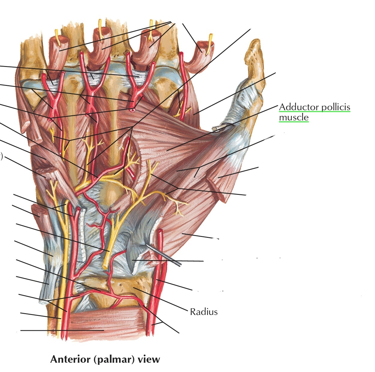
• Fig2 adductor pollicis muscle in the palm, Frank H. Netter, Atlas of Human Anatomy (7th edition) plate 455
• Fig3 Prosection 3 – The interossei and adductor pollicis of the hand. teach me anatomy website, https://teachmeanatomy.info/upper-limb/muscles/hand/
• FIG1 JOSEPH E. MUSCOLINO THE MUSCULAR SYSTEM MANUAL The Skeletal Muscles of the Human Body FOURTH EDITION FIGURE 9-14 Anterior view of the right adductor pollicis. PAGE 424
• Fig2 adductor pollicis muscle in the palm, Frank H. Netter, Atlas of Human Anatomy (7th edition) plate 455
• Fig3 Prosection 3 – The interossei and adductor pollicis of the hand. teach me anatomy website, https://teachmeanatomy.info/upper-limb/muscles/hand/
