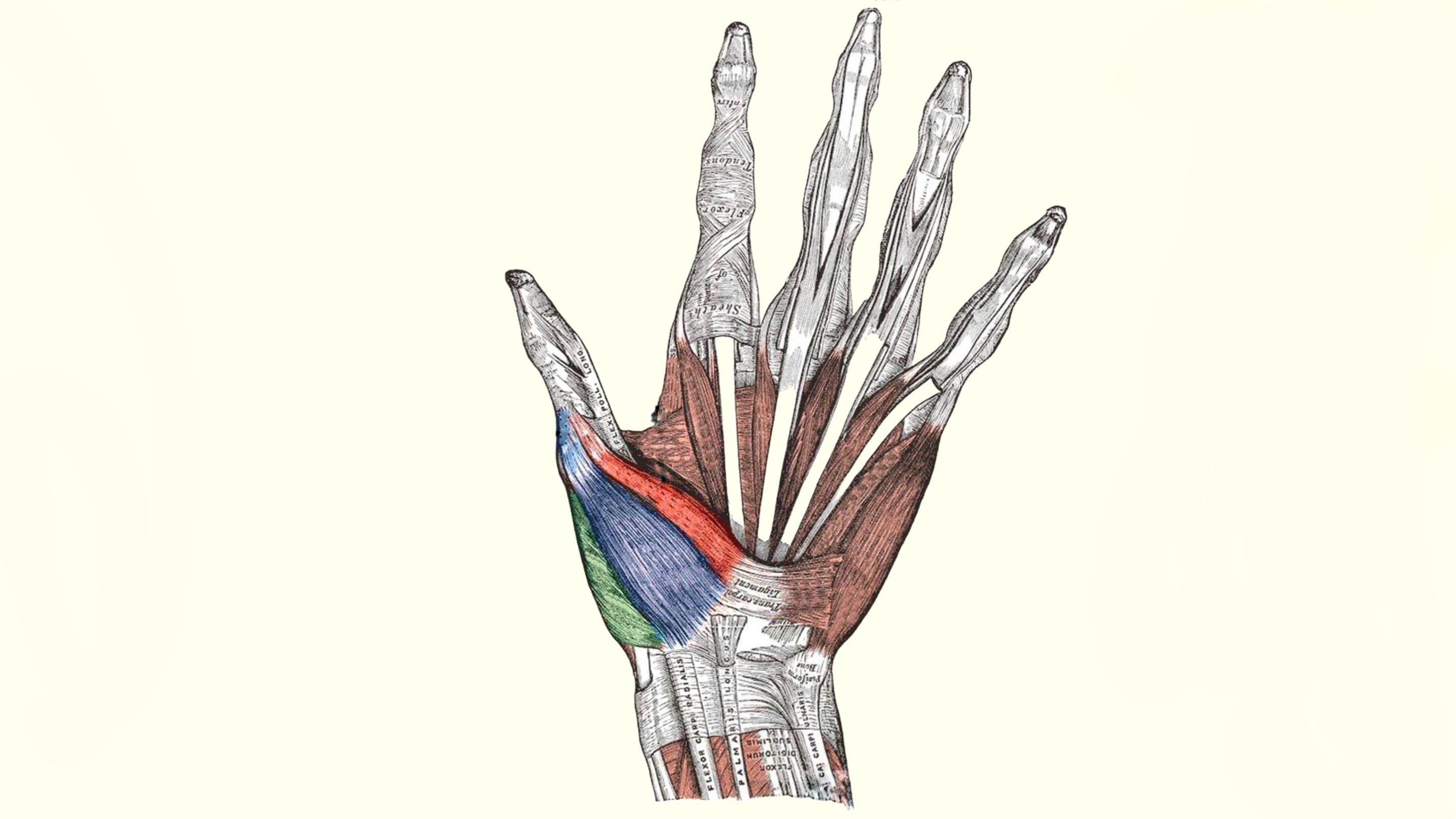
Opponens Pollicis Muscle
By : Omar M. Subhi Altaie• NAME
The first part of the name indicates that the main action of this muscle is opposition (which is moving the finger to touch other fingers), and the second part indicates that it applies that action to the thumb finger "pollicis".
• GENERAL
The opponens pollicis is one of three thenar muscle-group that are Located laterally on the palm side of the hand, it is a quadrangular muscle that lies deepest among the three in contact with the bone.
The most significant movement of the thumb is done by this muscle, we use opposition when picking up things. It applies that action and others on the saddle joint of the thumb ( carpometacarpal joint or CMC).
The most significant movement of the thumb is done by this muscle, we use opposition when picking up things. It applies that action and others on the saddle joint of the thumb ( carpometacarpal joint or CMC).

• SUPPLY
- The nerve supply of Opponens pollicis muscle is by the recurrent branch of the median nerve ( C8, T1) and in some cases by the deep terminal branch of the ulnar nerve.
- The blood supply of Opponens pollicis muscle is by the superficial palmar branch that originates from the radial artery. And there is additional blood supply might come from several other arteries such as :
• Radialis indicis artery
• Deep palmar arch
• Princeps pollicis artery
- The blood supply of Opponens pollicis muscle is by the superficial palmar branch that originates from the radial artery. And there is additional blood supply might come from several other arteries such as :
• Radialis indicis artery
• Deep palmar arch
• Princeps pollicis artery
● ORIGIN AND INSERTION
◇ Origin
Arises from Flexor retinaculum and tubercle of trapezium scaphoid of carpal bones as well as some of the abductor pollicis longus tendon.
◇ Insertion
The muscle fibres are coarse distally to reach the Anterior surface and some of the Lateral sides in the middle first metacarpal bone, as well as the base of the proximal phalanx of the thumb.

• Movement and action
The main action of opponens pollicis is the position which occurs in the muscle in what is called the triplanar oblique plane motion which is a combination of three plane actions that occur at the carpometacarpal joint of the thumb, which when bringing the pad (rounded palmar surface of the thumb) against the pad of another finger. Opposition involves several actions of the thumb like flexion , medial rotation , and abduction of the metacarpal of the thumb.
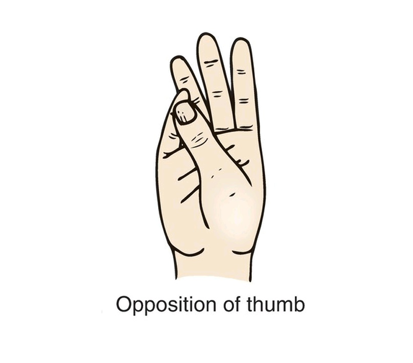
The opponens pollicis crosses the first carpometacarpal joint of the thumb from the medial side to attach to the metacarpal bone of the thumb when to muscle contracts it applies its other function which is Flexion, and that can be seen when the metacarpal bone of the thumb is pulled medially to the anterior to the palm flexing the Carpometacarpal joint of the thumb. And because it passes over the Metacarpophalgeal joint at the medial side so it flexes the proximal phalanx of the thumb too. A good piece of information to add is that when the muscle is flexing at the latter joint it also medially rotating the thumb because of the morphology of the articular surface of the joint, so that other actions that this muscle is capable of doing, the motion is coupled with the flexion; when moving the thumb toward the little finger with slightly changing its direction.
It also has another action which is ABduction ; when the fibres that cross the joint anteriorly and slightly medially, they pull the metacarpal bone of the thumb to move it to a position when it is vertical at a right angle with the surface of the palm .
It also has another action which is ABduction ; when the fibres that cross the joint anteriorly and slightly medially, they pull the metacarpal bone of the thumb to move it to a position when it is vertical at a right angle with the surface of the palm .
• Reverse action
As we know the normal action of any muscle is when the proximal attachment of the origin of that muscle is fixed and the distal attachment which is the insertion is the movable portion, but when the opposite of that happens we call it Reverse action.
The opposite of any of the actions that this muscle is capable of doing is required the fixation of the metacarpal bones due to some particular reason and moving the carpal bones of origin toward them and that makes the muscle contract.
The opposite of any of the actions that this muscle is capable of doing is required the fixation of the metacarpal bones due to some particular reason and moving the carpal bones of origin toward them and that makes the muscle contract.
• ANTAGONIST FUNCTIONS
• It slows reposition (opposite of opposition) of the thumb at the same joint
• Because it can do flexion so it slows the extension of the carpometacarpal joint of the thumb
• slows lateral rotation (due to the medial rotation action ) the metacarpal and medial rotation of the trapezium at the carpometacarpal joint of the thumb
• Because abduction that it can do so it slows the adduction of the carpometacarpal joint of the thumb.
• Examination
Ask the patient to oppose his thumb by making
" O " shape with his thumb against others fingers, and putting resistance on the thumb by not letting him do that action, if he can that easily, his opponens pollicis is intact.
" O " shape with his thumb against others fingers, and putting resistance on the thumb by not letting him do that action, if he can that easily, his opponens pollicis is intact.
• CLINICAL NOTES
The carpal tunnel syndrome happens after swelling of any of the contents of the tunnel like inflammation of synovial sheaths or reduction of the size of the tunnel; which will put compression on the median nerve and thus paralysis of the muscles that it supplies.
Opponens pollicis supplied by the median nerve, in this case, is paralyzed; the patient is not able to button his shirt or write text messaging on the phone.
A noticeable pain of the hand at the base of the thumb and down into the inside of the wrist may best describe one of the symptoms of injury.
To fix that condition of the compression and the symptoms that happened after, partial or complete surgical division of the flexor retinaculum is necessary; it is called carpal tunnel release surgery .
• Relations
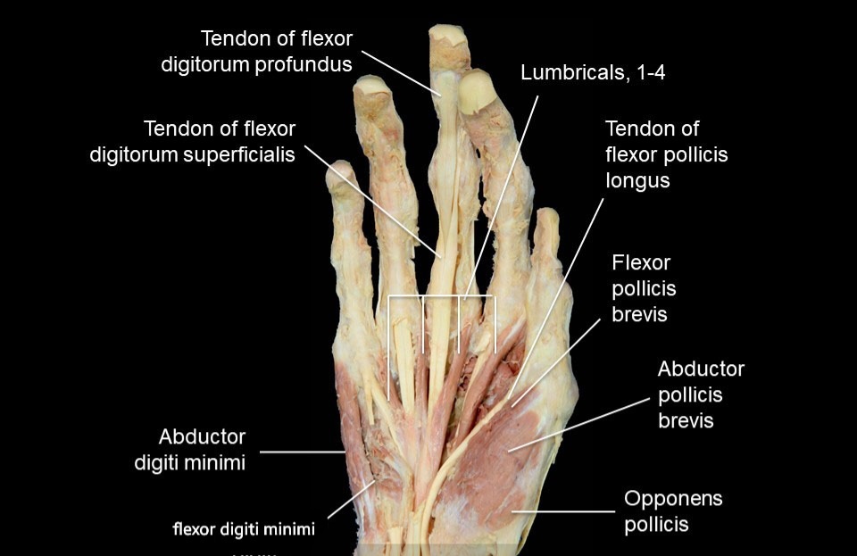
Opponens pollicis is one of the thenar eminence muscles on the lateral side of the hands.
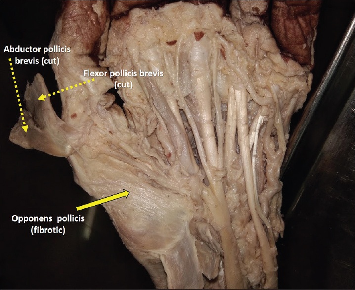
It is a short muscle that is superficial to it is the abductor pollicis brevis and medial to it, is the flexor pollicis brevis. The superficial head of flexor pollicis brevis is more often blended with opponens pollicis muscle.
It is a short muscle that is superficial to it is the abductor pollicis brevis and medial to it, is the flexor pollicis brevis. The superficial head of flexor pollicis brevis is more often blended with opponens pollicis muscle.
● REFERENCES
• JOSEPH E. MUSCOLINO THE MUSCULAR SYSTEM MANUAL The Skeletal Muscles of the Human Body FOURTH EDITION 391, 402-403
• Keith L. Moore, Arthur F. Dalley, Anne M. R Clinically Oriented Anatomy (7th Edition) page 777
• Snell's clinical anatomy by Region's (10th edition) pages 324-325, 424
• opponens pollicis physiopedia website https://www.physio-pedia.com/Opponens_polisis
• Opponens pollicis muscle Author: Roberto Grujičić MD • Reviewer: Gordana Sendić MD kenhub website https://www.kenhub.com/en/library/anatomy/opponens-pollicis-muscle
● IMAGES REFERENCES
• Cover image from https://twitter.com/Radford_DPT/status/965657989691650051?t=EkP55hkCYQb8S7DURp_NdQ&s=19
• Fig1 JOSEPH E. MUSCOLINO THE MUSCULAR SYSTEM MANUAL The Skeletal Muscles of the Human Body FOURTH EDITION page 403
• Fig2 Frank H. Netter, Atlas of Human Anatomy (7th edition) plate 452
• Fig3 Various positions of the hand and movements of the thumb. . (H) Anterior views showing movements of the thumb Figure 3.81. Snell's clinical anatomy by Region's (10th edition)page 423
• Fig4 (Figure 1): Dissection of the left hand showing fibrotic opponens pollicis after reflecting abductor and flexor pollicis brevis journal of current researches in scientific medicine website https://www.jcrsmed.org/viewimage.asp?img=JCurrResSciMed_2019_5_1_62_260643_f1.jpg
• Fig5 Prosection 2 – The lumbricals of the hand. Suárez-Quian & Vilensky. All in One Anatomy Review - Volume 1: Back and Upper Limb teach me anatomy website, https://teachmeanatomy.info/upper-limb/muscles/hand/
