The superficial veins of the upper limb
By : Ma'ab Maladh
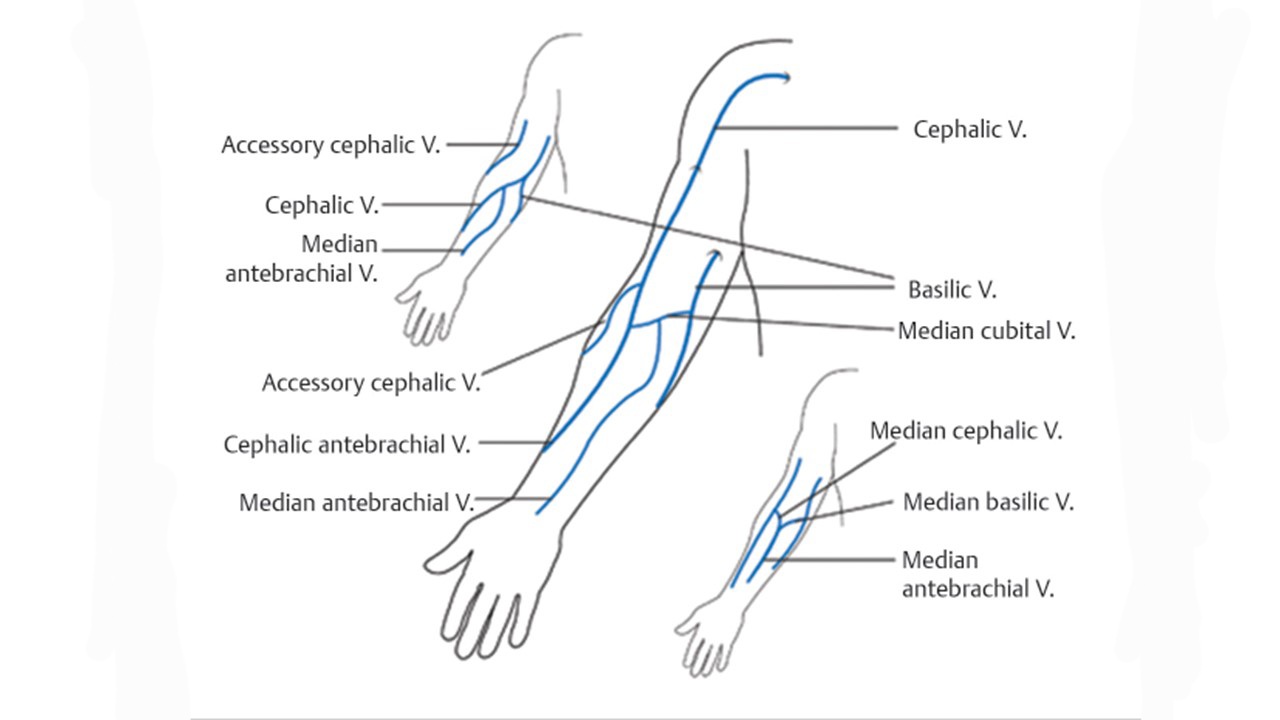
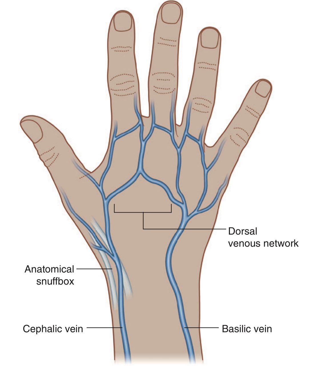
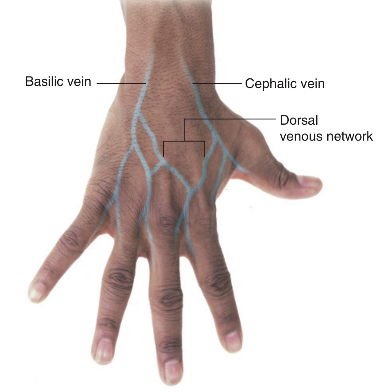
The superficial veins which lies in the superficial fascia drain into a dorsal venous network (Fig.1, Fig.2) on the back of the hand over the metacarpal bones, there are the basilic vein and the cephalic vein .
Like the arteries, the smooth muscle in the wall of the veins is innervated by sympathetic postganglionic nerve fibers that provide vasomotor tone. The origin of these fibers is similar to those of the arteries.
Note
A network of veins in the superficial fascia of the dorsum of the hand (back of the hand), formed by the dorsal metacarpal veins gives the origin of the basilic vein and the cephalic vein.Note
Like the dermatomal pattern, the logic for naming the main superficial veins of the upper limb cephalic (toward the head) and basilic (toward the base) becomes apparent when the limb is placed in its initial embryonic position.Like the arteries, the smooth muscle in the wall of the veins is innervated by sympathetic postganglionic nerve fibers that provide vasomotor tone. The origin of these fibers is similar to those of the arteries.
The cephalic vein
The draining area:
Drains the posterior and the lateral parts (the radial parts) of the hand, the forearm and the arm.
Course and relation:
arise from the lateral side of the dorsal venous network and passes over the anatomical snuffbox into the forearm. it ascends in the subcutaneous tissue (in the superficial fascia) in the dorsolateral aspect of the distal two thirds of the forearm.
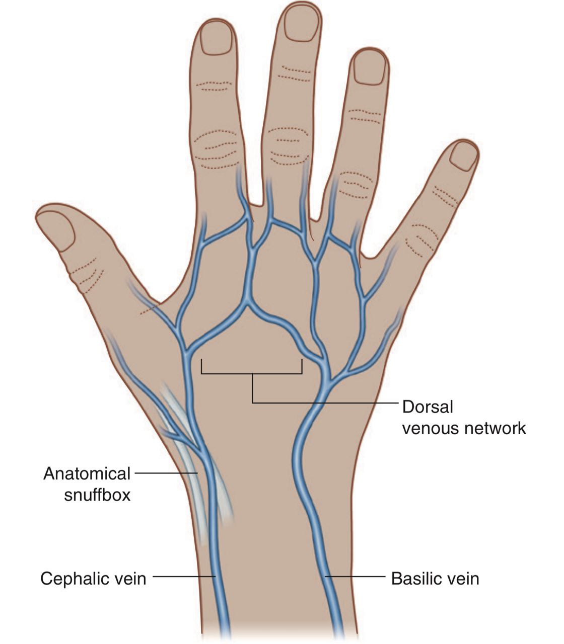

Fig.1 The dorsal venous network
Then it becomes anterolateral in the proximal third of the forearm few centimetres below elbow, runs over the bicipital aponeurosis and joins the basilic vein with the median cephalic vein (Fig.4) or with the median cubital vein (Fig.3).
Note
A sheet of connective tissue runs from the medial side of the bicipital tendon then merge the antebrachial fascia (the deep fascia of the forearm) covering the flexor muscles in the medial side of the forearm. It’s important to protect the structures laying under it in the cubital fossa (such as the brachial artery and the median nerve) and to lower the pressure of the bicipital tendon on the radial tuberosity ( the insertion of the biceps brachii) during pronation and supination (Fig.5).
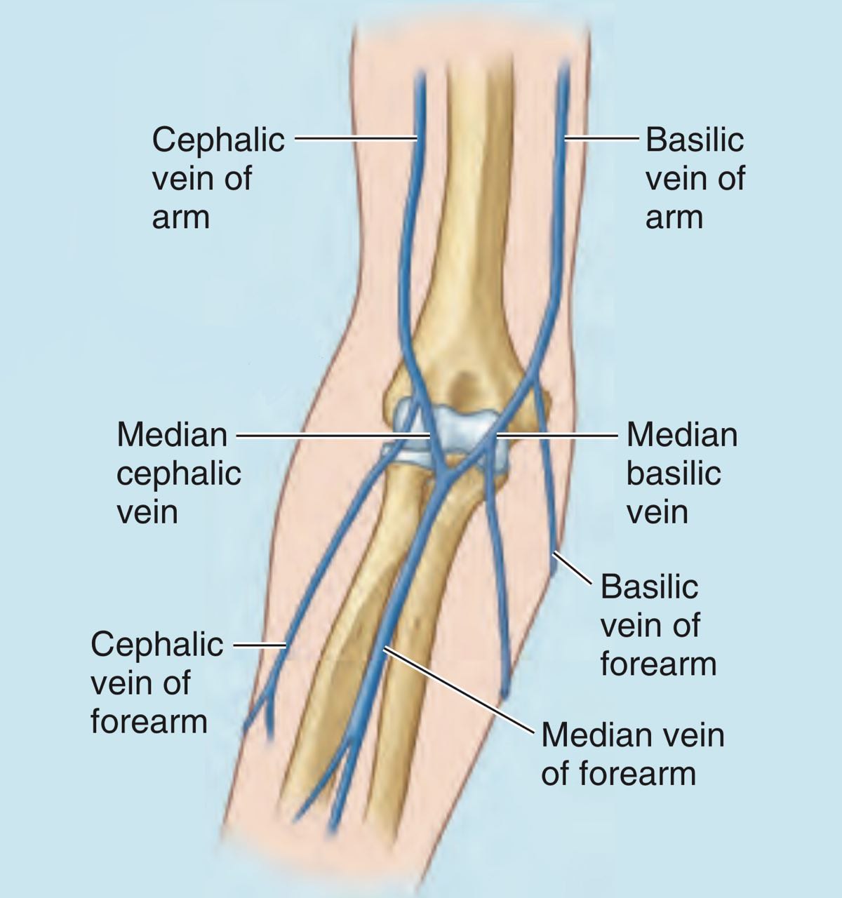
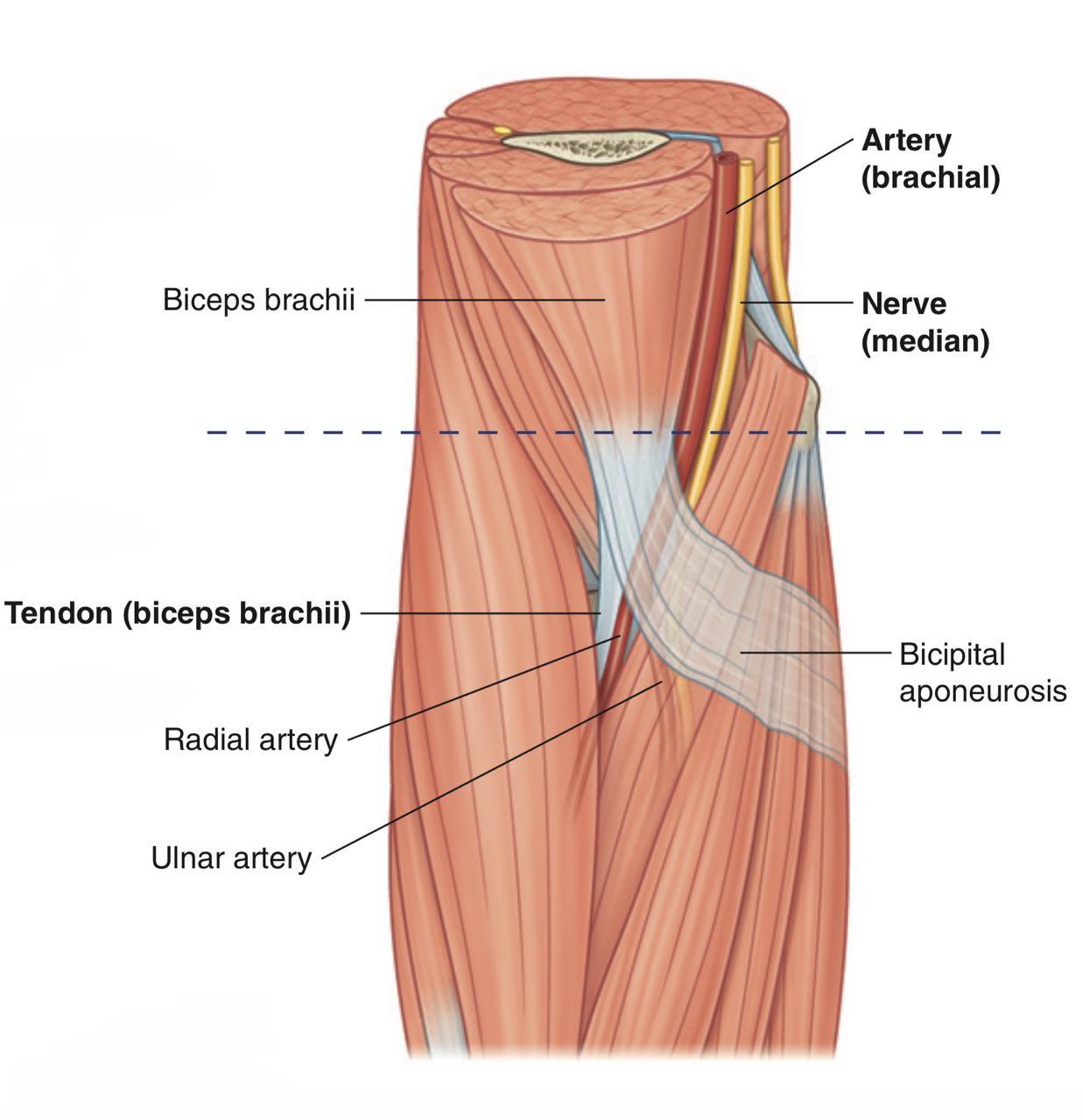
Fig.3 The median cubital vein
It ascends in the arm leteral to the biceps brachii muscle.
In the area of shoulder the cephalic vein passes into the inverted triangular cleft (the clavipectoral triangle) and courses superiorly between the deltoid muscle, pectoralis major muscle and clavicle along the deltopectoral groove . (fig.6)
In the superior part of the clavipectoral triangle , the cephalic vein passes deep to the clavicular head of the pectoralis major muscle and pierces the clavipectoral fascia to join the axillary vein . (Fig.7)
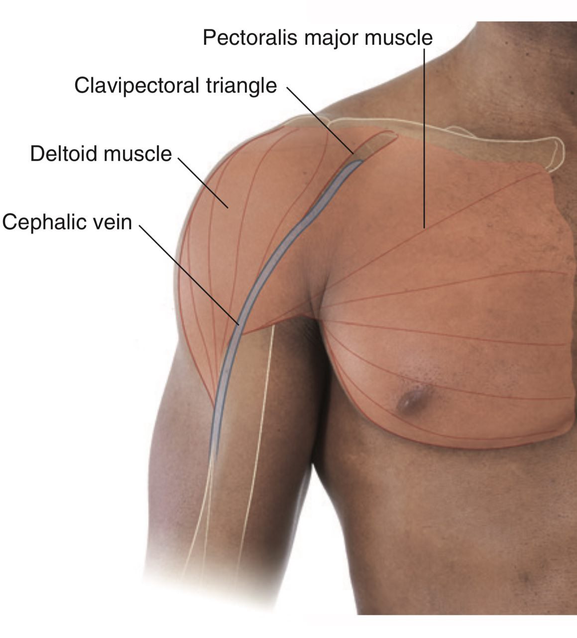

Fig.6 The deltopectoral groove
The basilic vein
The draining area:
drains the medial part (ulnar part) of the hand, forearm and arm.
Course and relation:
arise from the medial end of the dorsal venous network as a continuation. It ascends in the subcutaneous tissue ( in the superficial fascia) in the dorsomedial aspect of the distal two thirds of the forearm.
drains the medial part (ulnar part) of the hand, forearm and arm.
Course and relation:
arise from the medial end of the dorsal venous network as a continuation. It ascends in the subcutaneous tissue ( in the superficial fascia) in the dorsomedial aspect of the distal two thirds of the forearm.
Then it becomes anteromedial in the proximal third of the forearm few centimetres below the elbow, over the bicipital aponeurosis
Histologically aponeuroses have a role similar to tendons, they both attaches muscles to bones, but they differ in: the aponeurosis:
1-aponeurosis is made of layers of delicate, thin sheath.
2-function :
*absorbing energy while muscle contraction like spring and then returning it when they recoil.
*act as shock absorber.
*bears extra pressure and tension when muscle contracts.
3-it’s very rare for the aponeurosis to get injured.
the tendon:
1-tendons are tough and rope like.
2-function:
*allows attachment of the muscle to bone in it’s origin and insertion.
*provide strength and support that allow muscle to contract.
3-tendons get injured easily. joins the cephalic vein with the median cubital vein (Fig.3). Sometimes it joins the cephalic vein with the median basilic vein (Fig.4).
Note
A sheet-like deep fascia of pearly-white fibrous connective tissue containing fibroblasts (collagen-secreting spindleshaped cells) and bundles of collagenous fibres distributed in regular parallel pattern which makes it resilient (elastic or flexible),attaches the sheet-like muscles needing a wide area of attachment (such as: anterior abdominal aponeurosis(external oblique aponeurosis) (Fig.8), palmar (Fig.9) and planter aponeurosis (Fig.10)) Histologically aponeuroses have a role similar to tendons, they both attaches muscles to bones, but they differ in: the aponeurosis:
1-aponeurosis is made of layers of delicate, thin sheath.
2-function :
*absorbing energy while muscle contraction like spring and then returning it when they recoil.
*act as shock absorber.
*bears extra pressure and tension when muscle contracts.
3-it’s very rare for the aponeurosis to get injured.
the tendon:
1-tendons are tough and rope like.
2-function:
*allows attachment of the muscle to bone in it’s origin and insertion.
*provide strength and support that allow muscle to contract.
3-tendons get injured easily.
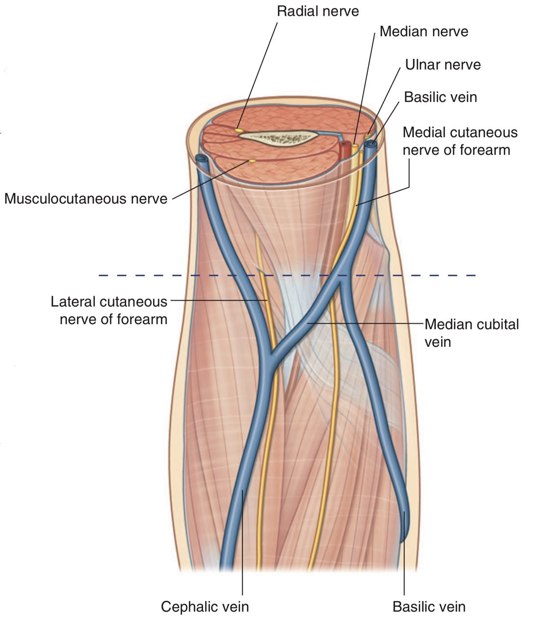

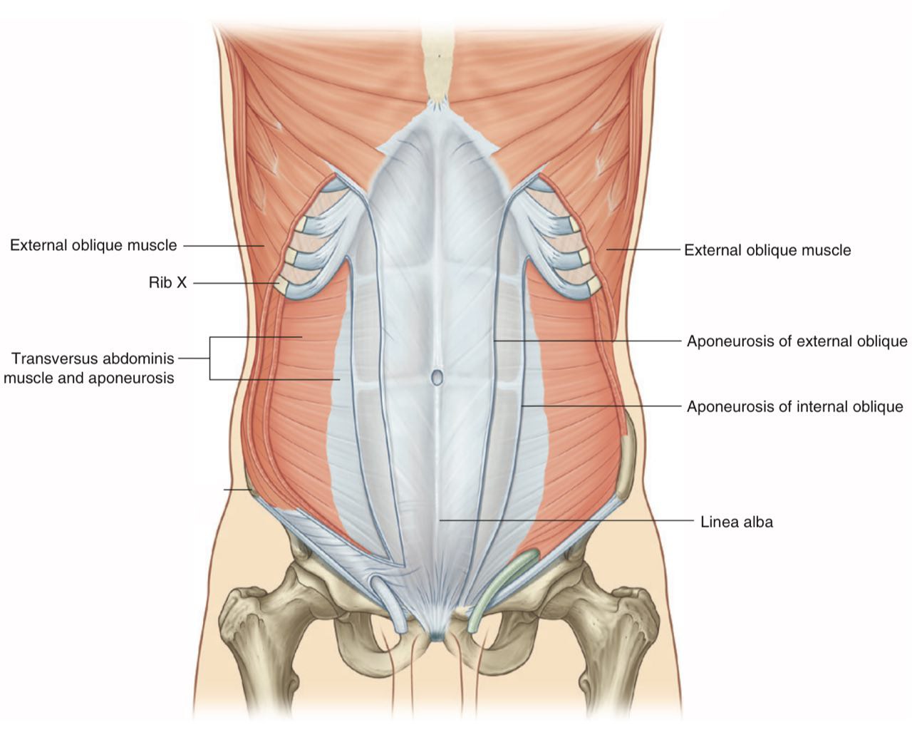
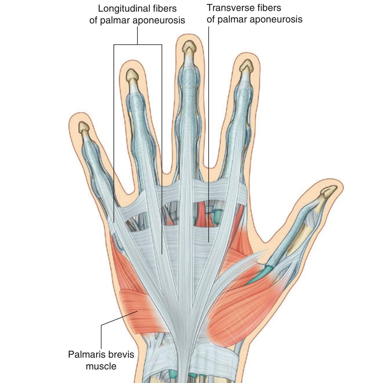

Fig.3 The median cubital vein
It ascends vertically in the arm medial to the biceps , and in the halfway up the arm (in the middle of the arm) it piece (enter) the brachial fascia (deep fascia of the arm).
At the lower border of teres major the two venae comitantes of the brachial artery join it. Then the Basilic vein becomes axillary vein .
At the lower border of teres major the two venae comitantes of the brachial artery join it. Then the Basilic vein becomes axillary vein .

fig.11
Clinical notes
The superficial veins are clinically important and are used for venipuncture ( to test or monitor blood components, or to remove blood due the excess levels of iron or erythrocytes), transfusion ( like in hypovolemic shock when someone loose a lot of blood or body fluids like in diarrhea, throwing or sweating so the heart can’t get enough blood and starts to loose it’s function), and cardiac catheterisation.
Usually in venipuncture and transfusion the median cubital vein is used. Other veins that can be used in the cubital fossa in cephalic vein , basilic vein , median antebrachial vein .
You should know where to obtain blood from the arm. When a patient is in a state of shock, the superficial veins are not always visible.
In the cubital fossa, the median cubital vein is separated from the underlying brachial artery by the bicipital aponeurosis. This is important because it protects the artery from the mistaken introduction into its lumen (cavity) of irritating drugs that should have been injected into the vein.
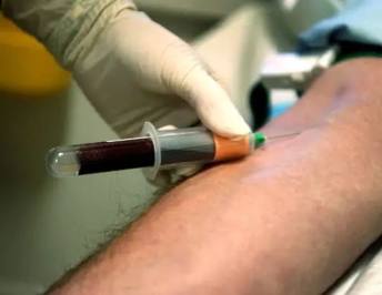
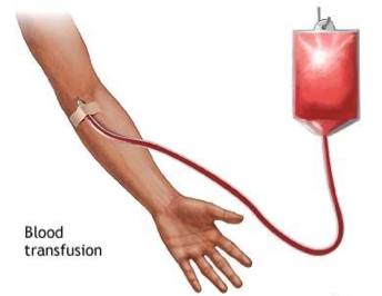
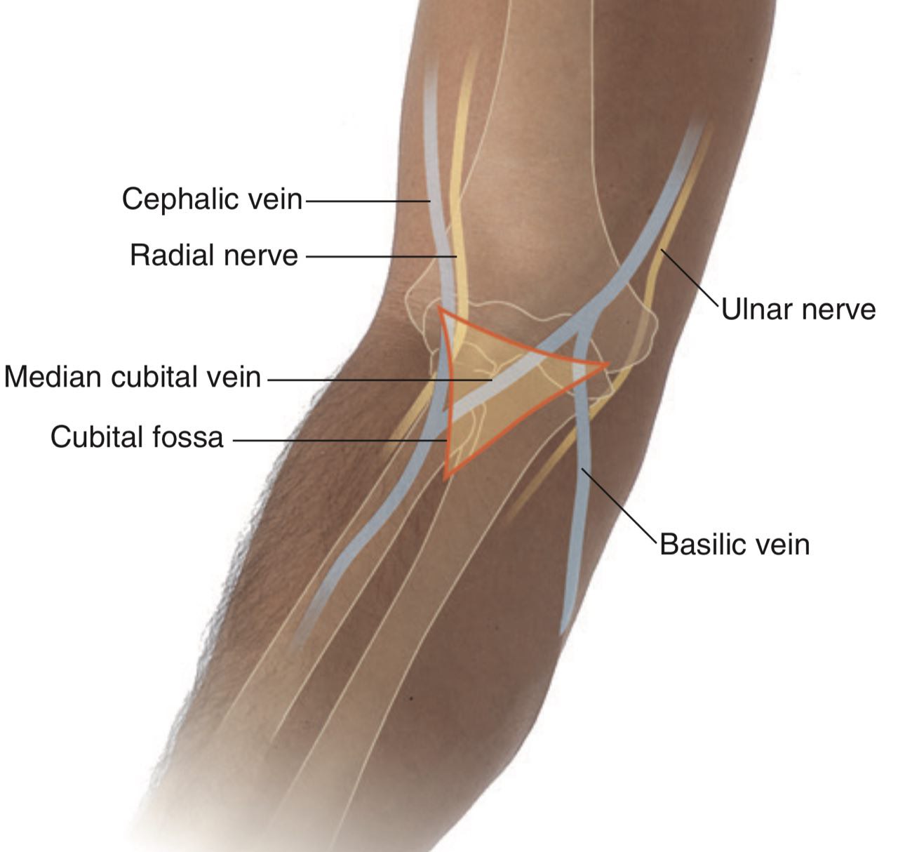
Fig.12 Venipuncture for blood test
Cardiac catheterization :
Cardiac catheterization is a procedure in which a thin, flexible tube (catheter) is guided through a blood vessel to the heart to diagnose or treat certain heart conditions, such as clogged arteries or irregular heartbeats.
The median basilic or basilic veins are good choice for central venous catheterization , because from the cubital fossa until the basilic vein reaches the axillary vein, the basilic vein increases in diameter and is in direct line with the axillary vein.
The valves of the axillary vein may be a troublesome but abduction of the shoulder will permit the catheter to pass the obstacle.
We don’t choose the cephalic vein because
1) it doesn’t increase in diameter as it ascends the arm.
2) it frequently divide into smaller branches as it lies in the deltopectoral triangle.
3) it drains into the axillary vein at right angle and it is difficult to insert the catheter around this angle
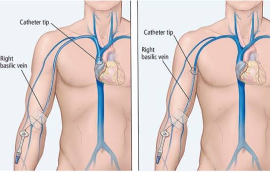
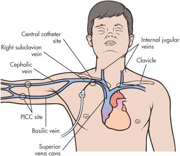

Fig.15 Cardiac catheterization
References
1. Moore, K. L., Dalley, A. F., & Agur, A. M. (2014). Clinically oriented anatomy 7th edition,S.689-691,743,744
2. Drake, R., Vogl, A. W., Mitchell, A. W., Tibbitts, R., & Richardson, P. (2020). Gray's Atlas of Anatomy E-Book. Elsevier Health Sciences, S.688,725,748,805.
3. Snell, R. S. (2012). Snell's Clinical Anatomy by Regions. 9th South Asian ed.,S.369,370
4. Gosling, J. A., Harris, P. F., Humpherson, J. R., Whitmore, I., & Willan, P. L. (2008). Human anatomy, color atlas and textbook E-Book. Elsevier Health Sciences.
5. The cephalic vein and the basilica vein ,Shahab Shahid ,kenhub
Image references:
1. Moore, K. L., Dalley, A. F., & Agur, A. M. (2014). Clinically oriented anatomy 7th edition,Fig. B6.18.
2. Drake, R., Vogl, A. W., Mitchell, A. W., Tibbitts, R., & Richardson, P. (2020). Gray's Atlas of Anatomy E-Book. Elsevier Health Sciences, Fig.2.42, 4.31, 6.115, 7.67, 7.51, 7.77.B., 7.77.D., 7.99, 7.112, 7.121.C., 7.123.B.
3. Gosling, J. A., Harris, P. F., Humpherson, J. R., Whitmore, I., & Willan, P. L. (2008). Human anatomy, color atlas and textbook E-Book. Elsevier Health Sciences.Fig. 3.26.
4. Frank, H. N. (2018). Netter Atlas of Human Anatomy 7th Ed 2018.plate 406.
