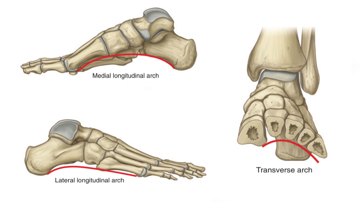
The Arches of Foot
By : Hafsa OmarDefinition
Arches of foot are made of tarsal and metatarsal bones, these bones are connected to ligament which give flexibility to the foot.
Tarsal and metatarsal bones are arranged in the shape of arches, these arches are:
1.Medial longitudinal arch
2.Lateral longitudinal arch
3.Transverse arch
Tarsal and metatarsal bones are arranged in the shape of arches, these arches are:
1.Medial longitudinal arch
2.Lateral longitudinal arch
3.Transverse arch
Function
1.Distributing the weight of the body over the foot.
2.Absorbing the shocks (forces) that result through moving.
3.Giving the foot the ability to adapt to changes in surface contour.
Note: these arches become slightly flattened by body weight during standing and return curvature when the weight is removed.
2.Absorbing the shocks (forces) that result through moving.
3.Giving the foot the ability to adapt to changes in surface contour.
Note: these arches become slightly flattened by body weight during standing and return curvature when the weight is removed.
Supporters of the arches
1.Shape of the bones that form the arches.
2.Ligaments.
3.Muscles (especially tibialis anterior, tibialis posterior and fibularis longus muscles).
All of these play an important role in maintaining and supporting the arches of foot.
Note: tibialis anterior m., fibularis longus m. and small muscles of foot are inactive in the static position, but during walking and running these muscles become active and support the arches.
Medial longitudinal arch
It is higher than lateral longitudinal arch.
It runs at the medial aspect of the foot.
It consists of:
1.Calcaneus
2.Talus
3.Navicular
4.Three cuneiforms
5.Medial three metatarsal bones
6.Sesamoid bones (two sesamoid bones under the head of 1st metatarsal bone)
It runs at the medial aspect of the foot.
It consists of:
1.Calcaneus
2.Talus
3.Navicular
4.Three cuneiforms
5.Medial three metatarsal bones
6.Sesamoid bones (two sesamoid bones under the head of 1st metatarsal bone)
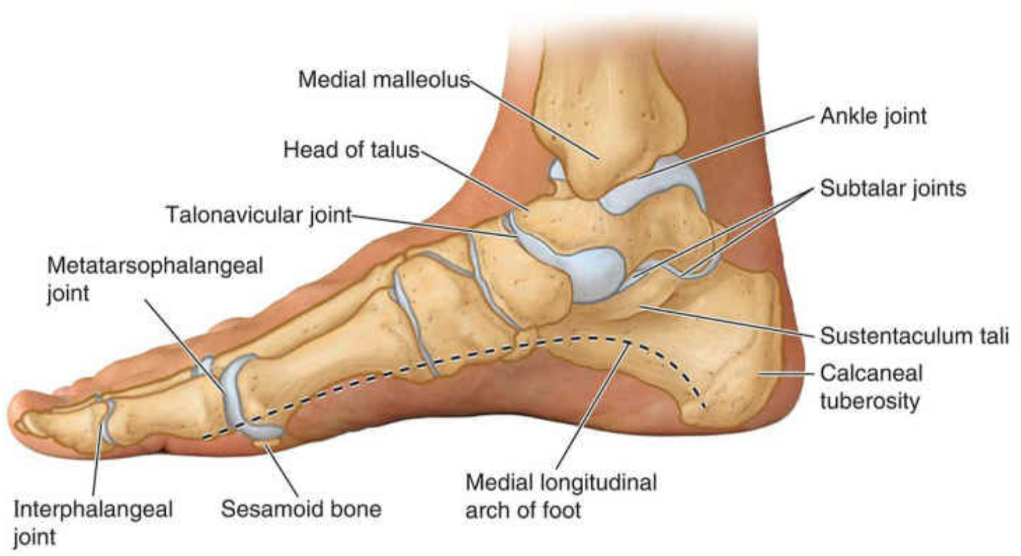
Medial longitudinal arch
Muscles that support medial longitudinal arch:
1.Tibialis anterior and posterior
2.Flexor digitorum longus and brevis (both medial parts)
3.Flexor hallucis longus and brevis
4.Abductor hallucis
Ligament that support medial longitudinal arch:
1.Plantar aponeurosis (medial half)
2.Spring ligament (or plantar calcaneonavicular ligament)
3.Long plantar ligament (between the calcaneus and the cuboid bones, it continues to the metatarsal bones)
4.Short plantar ligament (or plantar calcaneocuboid ligament)
5.Medial ligament of the ankle joint

Ligament that support medial longitudinal arch
Lateral longitudinal arch
It is flatter than the medial longitudinal arch and rest on ground during standing, because of the exerted pressure on the ground by the lateral margin of the foot is great.
It consists of:
1.Calcaneus
2.Cuboid
3.Lateral two metatarsal bones
It consists of:
1.Calcaneus
2.Cuboid
3.Lateral two metatarsal bones
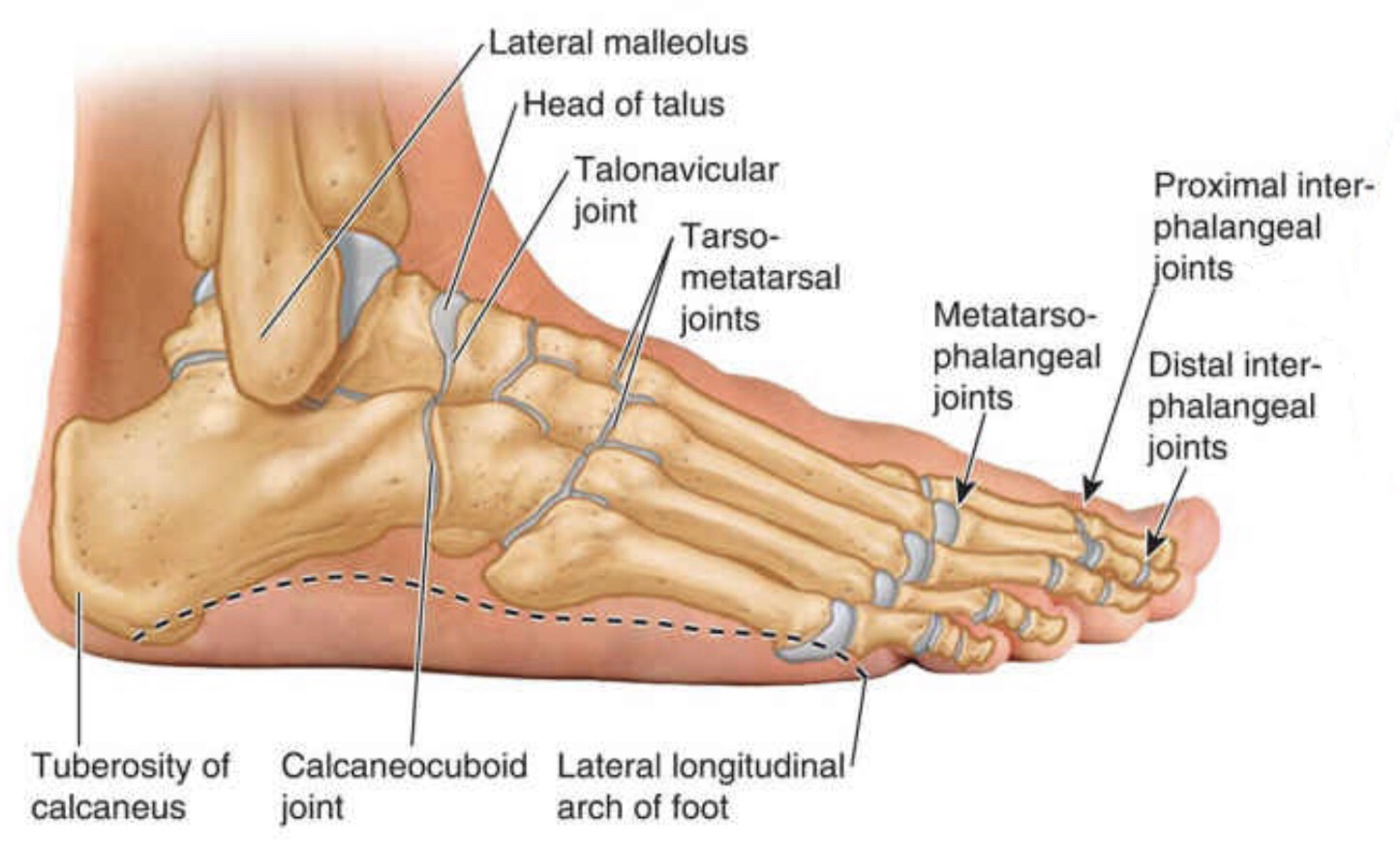
Lateral longitudinal arch
Muscles that support lateral longitudinal arch
1.Fibularis (peroneus) longus and brevis
2.Flexor digitorum longus and brevis (both lateral parts)
3.Abductor digiti minimi
Ligament that support lateral longitudinal arch
1.Plantar aponeurosis (lateral half)
2.Long plantar ligament
3.Short plantar ligament
4.Spring ligament
1.Fibularis (peroneus) longus and brevis
2.Flexor digitorum longus and brevis (both lateral parts)
3.Abductor digiti minimi
Ligament that support lateral longitudinal arch
1.Plantar aponeurosis (lateral half)
2.Long plantar ligament
3.Short plantar ligament
4.Spring ligament
Transverse arch
runs in the coronal plane of the foot.
It consists of:
1.Cuboid
2.Three cuneiform
3.Metatarsal bases (5)
Note: the medial and the lateral longitudinal arches acts as a pillar for the transverse arch and maintain it.
It consists of:
1.Cuboid
2.Three cuneiform
3.Metatarsal bases (5)
Note: the medial and the lateral longitudinal arches acts as a pillar for the transverse arch and maintain it.

Bones forming the transverse arch
Note: the tendons of tibialis posterior and fibularis longus muscles pass under the sole of foot as a stirrup help keeping the curvature of the transverse arch.
Muscles that support transverse arch:
1.Fibularis longus and brevis
2.Tibialis posterior
3.Dorsal interossei
4.Adductor hallucis (the transverse head)
Ligament that support transverse arch:
1.Plantar aponeurosis
2.Long plantar ligament
3.Short plantar ligament
3.Spring ligament
4.Deep transverse metatarsal ligaments
Clinical notes
1. Pes planus
Pes planus or (flat foot) is a condition of foot deformity involves the absence of medial longitudinal arch.
Pes planus may be congenital (present from birth) or acquired. Pes planus before 3 age is normal due to thick subcutaneous fat pad in the sole of foot, when the child get older the fat is loss and medial longitudinal arch becomes visible.
Pes planus can be:
1.Flexible (medial longitudinal arch disappears during carrying
weights but the arch becomes visible when the weight is removed).
2.Rigid (the foot remains flat even when the weight is
removed).
Pathogens of the pes planus:
1.Bone deformity: such as fusion of the tarsal bones
2.Acquired flat foot because of dysfunction of the tibialis posterior muscle due to trauma, degeneration with age (lose functional ability) or denervation (loss of nerve supply)
3.Ligament laxity
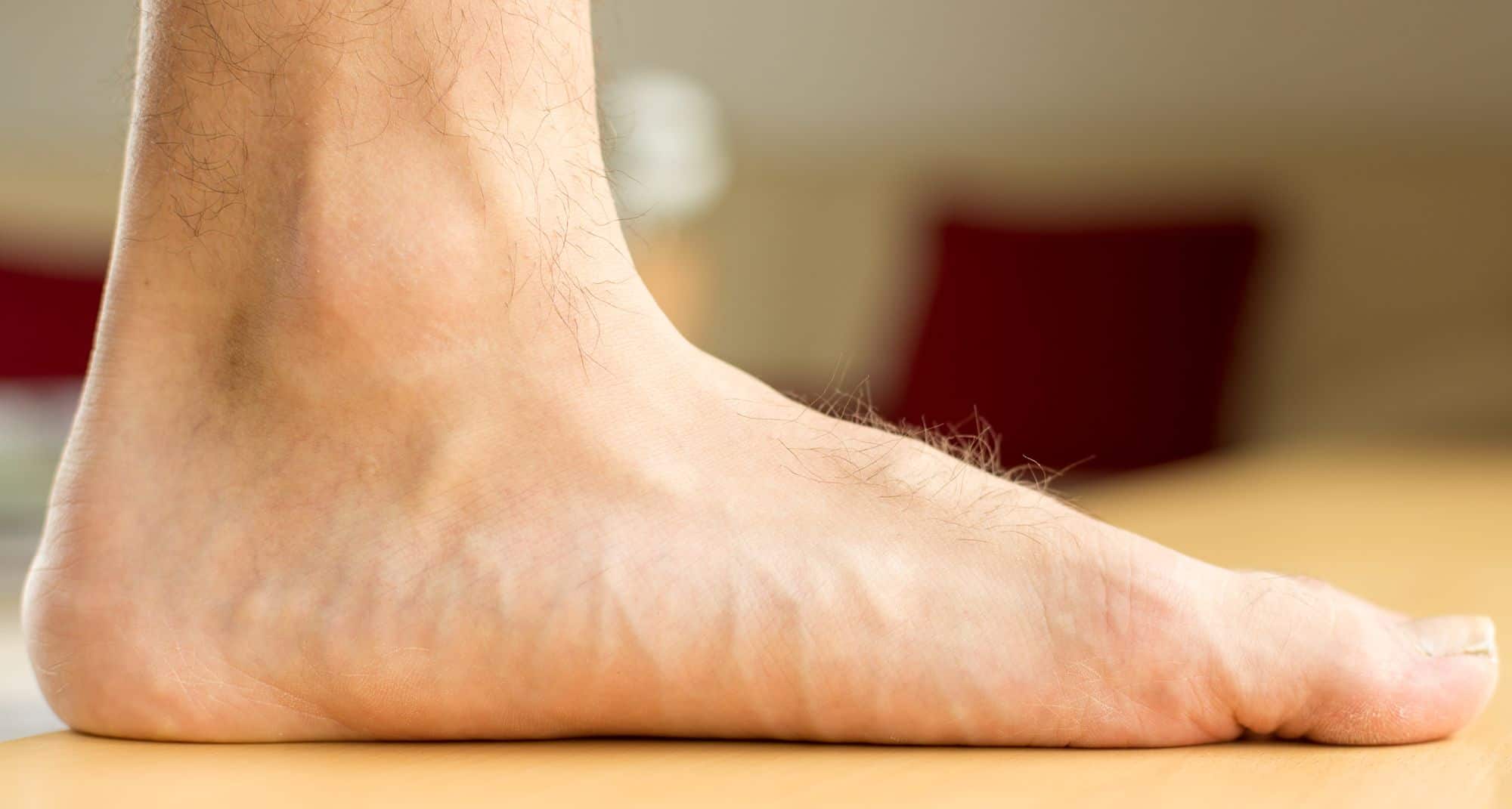
Pes planus
2. Pes cavus:
Pes cavus (or high arch) is a condition in which the medial longitudinal arch is higher than the normal.
It causes pressure on the heel and ball (the portion between the arch and toes) of foot at walking or standing.
It can occur at any age in one or both foot. Most cases are caused by muscle imbalance, in many cases it results from poliomyelitis.
Symptoms:
1. Claw toes
2. Pain
3. Tripping
4. Fractures
Treatment: involves wearing special shoes and using orthotics. Surgery may occur if other methods is not efficient.

Cavus foot
References
1- KEITH L. MOORE, ARTHUR F. DALLEY, ANNE M. AGUR, MOORE Clinically Oriented Anatomy (2018) EIGHTH EDITION, P/(1833-1834-1835-1836, 1864)
2- LAWRENCE E. WINESKI, SNELL'S CLINICAL ANATOMY BY REGIONS (2018) 10th edition, P/ (588-589)
3- Arches of the foot, Author: Charlotte O'Leary BSc, Kenhub
https://www.kenhub.com/en/library/anatomy/arches-of-the-foot
4- The Arches of the Foot, Author: Sam Little, TeachMe Anatomy https://teachmeanatomy.info/lower-limb/misc/foot-arches/
5- Pes Cavus, Author: Ezekiel Richardson, buoy health,
https://www.buoyhealth.com/learn/pes-cavus
References of images
1- Cover image: Gray’s Anatomy for Students, Fig. 6.113 (A,B,C).
2- Fig 1: Moore Clinically Oriented Anatomy, FIGURE 7.106.
3- Fig 2: Gray’s Anatomy for Students, Fig. 6.114
4- Fig 3: Moore Clinically Oriented Anatomy, FIGURE 7.106.
5- Fig 4: SNELL'S CLINICAL ANATOMY BY REGIONS, Figure 11.66
6- Fig 5: pes planus, LIMBIONICS, https://limbionics.com/blog/pes-planus-flat-feet/
7- Fig 6: cavus foot, Samarpan Physiotherapy Clinic,
https://samarpanphysioclinic.com/cavus-foot/amp/
