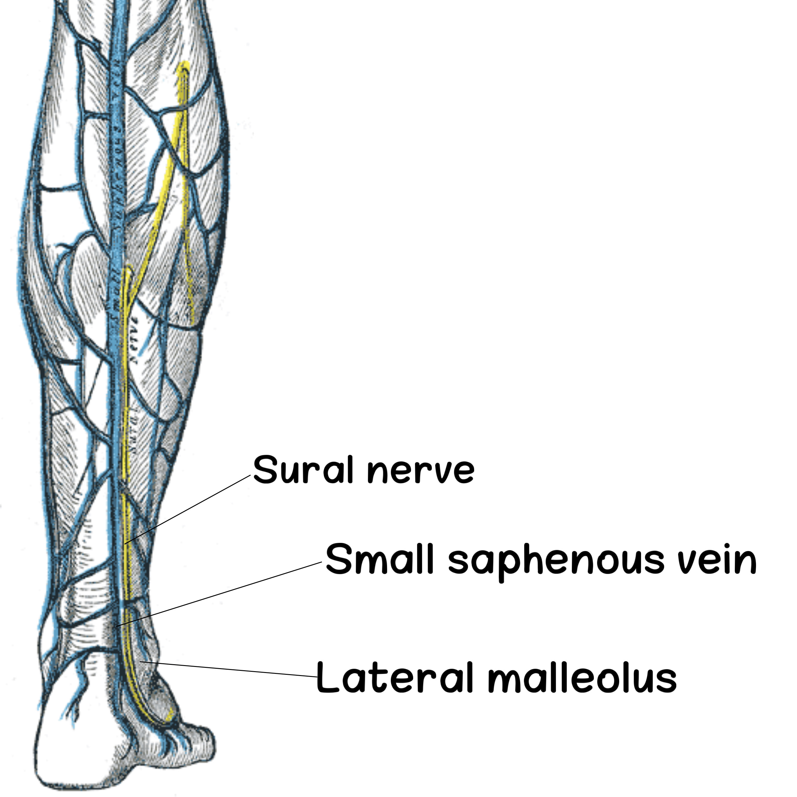
Structures That Pass Behind Lateral Malleolus Above Superficial Peroneal Retinaculum
By : Amna Mohammed1. Small saphenous vein
It is a continuation of the dorsal venous arch of the foot with dorsal vein of the lateral toe. It passes behind the lateral malleolus above the superficial peroneal retinaculum and ascends along the posterior aspect of the leg to end in the popliteal vein.
2. Sural nerve(S1-S2)
It is a cutaneous nerve.
It is formed by the union of the branch of tibial nerve and the branch of common fibular nerve .
It passes behind the lateral malleolus above the superficial peroneal retinaculum to supply the lateral side of the foot and the lateral toe.
It is formed by the union of the branch of tibial nerve and the branch of common fibular nerve .
It passes behind the lateral malleolus above the superficial peroneal retinaculum to supply the lateral side of the foot and the lateral toe.

Posterior view.
Right lower limb.
Clinical Note
The sural nerve has an important role at this region in local anesthesia of the led. Because of its long course and being superficial, it needs less injections and less quantity of anesthesia.
References
Moore-Clinically Oriented Anatomy (7th Edition)-538
Snell’s Clinical Anatomy By Regions (10th Edition)-1335,1347
Sural Nerve Block - Joshua J Solano -medscape- https://emedicine.medscape.com/article/83199-overview#a1
Henry Gray-Anatomy Of The Human Body (20th Edition)-582
Snell’s Clinical Anatomy By Regions (10th Edition)-1335,1347
Sural Nerve Block - Joshua J Solano -medscape- https://emedicine.medscape.com/article/83199-overview#a1
Henry Gray-Anatomy Of The Human Body (20th Edition)-582
