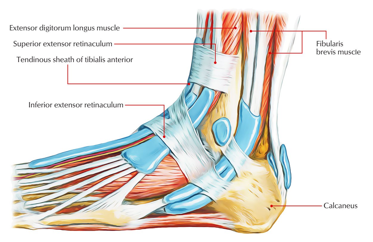
Extensor Retinaculum of foot
By : zahraa Al-ObaidiDefinition And Location
It is a strong fibrous band located at the distal end of the leg and specifically , in front of the ankle joint and the dorsal aspect of the foot.
Function
keeping the tendons(especially the extensor ones) in their position during the muscles contraction (which causes movement) , and also connecting the tibia and fibula bones.
figure no1
figure no1

There are two extensor retinacula in the foot:
1.Superior extensor retinaculum.
2.Inferior extensor retinaculum
2.Inferior extensor retinaculum
Superior extensor retinaculum
It is a horizontal band. It is attached:
Medially : to the anterior board of tibia.
Laterally : to the anterior board of fibula.
figure no2
Medially : to the anterior board of tibia.
Laterally : to the anterior board of fibula.
figure no2
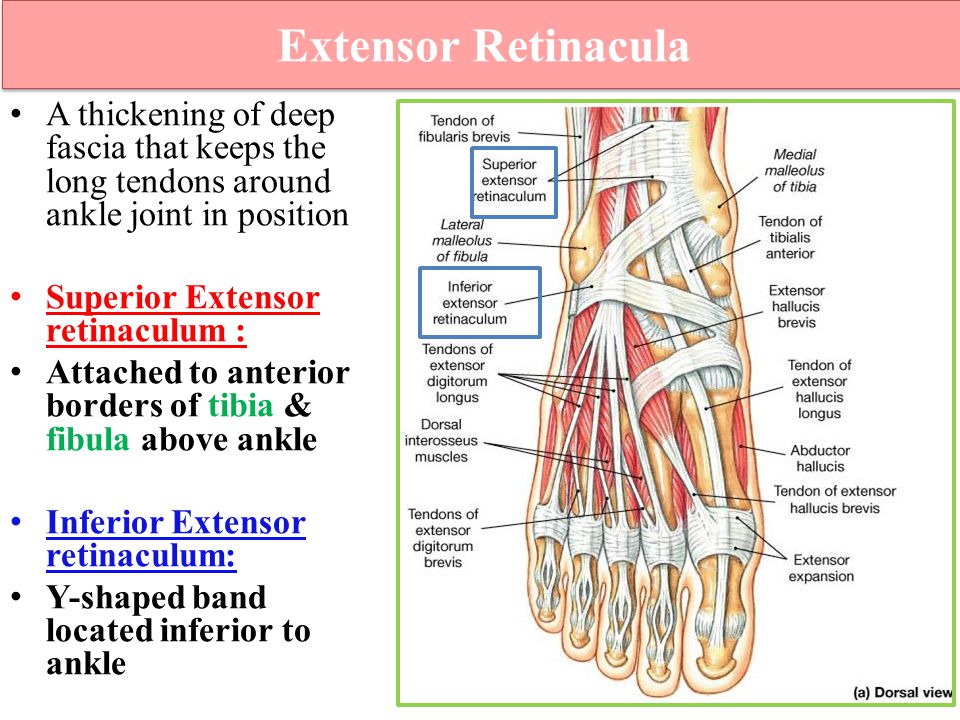
Inferior extensor retinaculum
It is a y-shaped band.
It is located in front of the ankle joint.
It consists of three parts:
1_Stem : it is attached to the superior surface of calcaneus.
2_Upper band : it is attached to the anterior board of the medial malleolus.
3_Lower band : it is attached to the deep fascia of the sole. It extends medially to fuse with the planter aponeurosis.
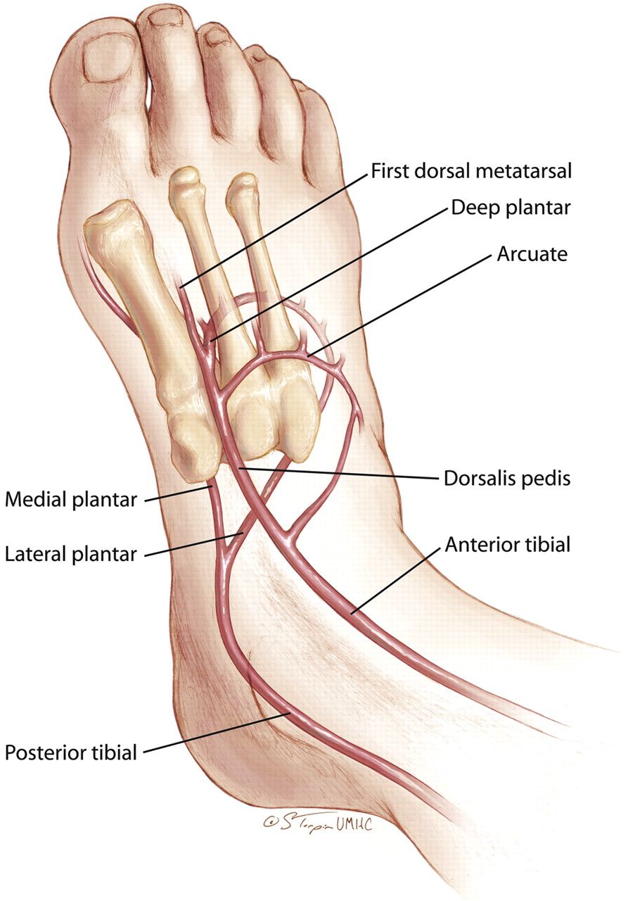
The structures that pass deep to the extensor retinaculum
( from medial to latera l)
•Tibialis anterior tendon
•Extensor hallucis longus tendon
•Anterior tibial vessels •Deep peroneal nerve(fibular nerve)
•Extensor digitorum longus tendon
•peroneus tertius tendon
Note : all these tendons are surrounded by synovial sheaths.
Note : all the structures that pass deep to the superior extensor retinaculum are the same structures that pass deep to the inferior extensor retinaculum, except the anterior tibial artery, it turn into dorsalis pedis artery at the inferior extensor retinaculum.
figure no3
The structures that pass superficial to the extensor retinaculum
( from medial to lateral)
•Superficial peroneal(fibular) nerve •Saphenous nerve
•Great saphenous vei
•Superficial peroneal(fibular) nerve •Saphenous nerve
•Great saphenous vei
Extensor tendonitis
All the tendons at the extensor retinaculum are surrounded by synovial sheaths so, any probable overpressure or stress , it will cause an inflammation which is called extensor tendonitis.
The most susceptible people to have extensor tendonitis are runners, dancers and skaters, because of their tightly tied shoes or because of an infection in their feet.
figure no4
The most susceptible people to have extensor tendonitis are runners, dancers and skaters, because of their tightly tied shoes or because of an infection in their feet.
figure no4

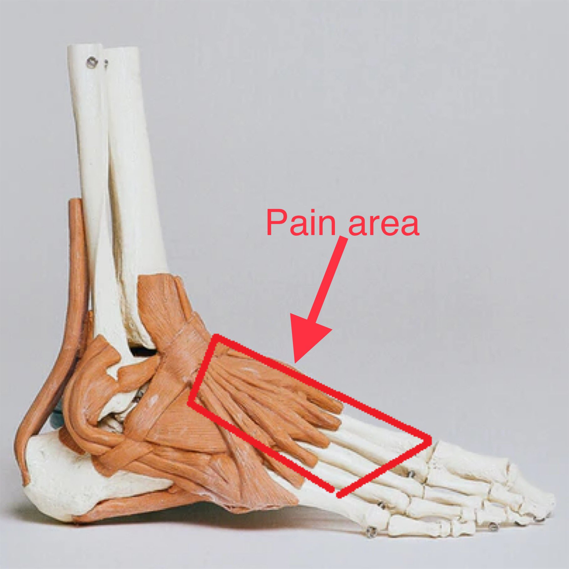
Symptoms
1.Pain in the front of the ankle or at the top of the foot, which increases during running and physical exercises.
2.Redness
3.Discoloration of the tendon
4.Warmth around the tendon 5.Bruising 6.Swelling
2.Redness
3.Discoloration of the tendon
4.Warmth around the tendon 5.Bruising 6.Swelling
Treatment
Here is the most important steps, the patient should do:
1.The most important procedure is taking a rest
2.Putting ice at the place of the pain for a while
3.Putting an elastic bandage and it shouldn't be tight
4.Taking anti-inflammatory drugs
1.The most important procedure is taking a rest
2.Putting ice at the place of the pain for a while
3.Putting an elastic bandage and it shouldn't be tight
4.Taking anti-inflammatory drugs
References
1. Henry Gray; Anatomy of the Human Body: Gray's Anatomy(1918); 20th edition;p.488
2. Keith L. Moore , Arthur F. Dalley A. M. R. Agur; Moore clinically oriented anatomy 7thedition;pp.610,619,688,750-752,757,589
3.Dr. Lawrence E. Wineski,PhD;SNELLIS CLINICAL ANATOMY BY REGIONS 10th edition; pp.1338-1339,1334,1351,1385
4. Extensor retinaculum (foot);Dr Maulik S Patel;radiopaedia; https://radiopaedia.org/articles/extensor-retinaculum-foot
5. Ankle Stability - Retinaculum Connection;Dr. Brian Abelson;KineticHealth;
https://www.kinetichealth.ca/post/2012/02/07/ankle-stability-therentinaculum
6. Extensor Tendinitis;ClevelandClinic;
https://my.clevelandclinic.org/health/diseases/23126-extensortendinitis#symptoms-and-causes
References of figures
interface figure: Superior Extensor Retinaculum – Earth's Lab
https://www.earthslab.com/anatomy/superior-extensor-retinaculum/
figure no2_Костно-фиброзные каналы и синовиальные влагалища кисти.(14-15 ...
https://cyberpedia.su/26xd9a5.html
figure no3Foot Anatomy Dr Rania Gabr. - ppt video online download
https://slideplayer.com/amp/4337995/
figure no4Dorsalis Pedis Artery - Stepwards
https://images.app.goo.gl/WmYkd7gmXXYGJCdb7
figure no 5What Causes Pain on Top of the Foot? — Dr. Elton
https://images.app.goo.gl/Cz662PCYt3byFQ3D8
Twitter 上的 Sports Physiotheray:"EXTENSOR TENDONITIS ...https://images.app.goo.gl/XabiMXA1cLVQ7VMD7
