


STRUCTURES THAT PASS ANTERIOR TO THE EXTENSOR RETINACULUM
By : Amna Mohammed
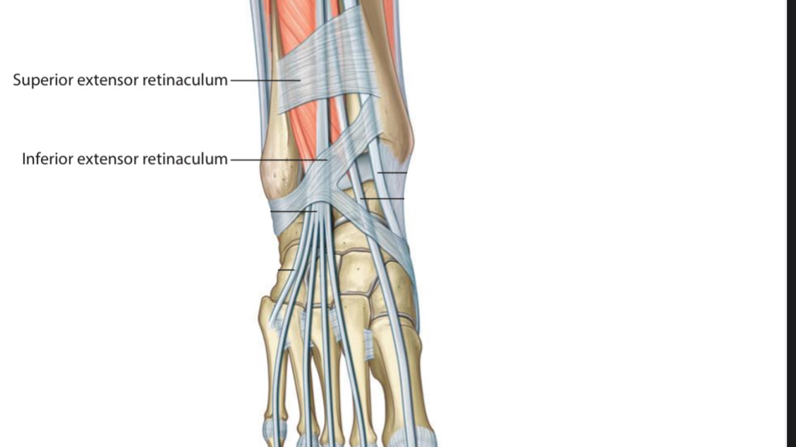
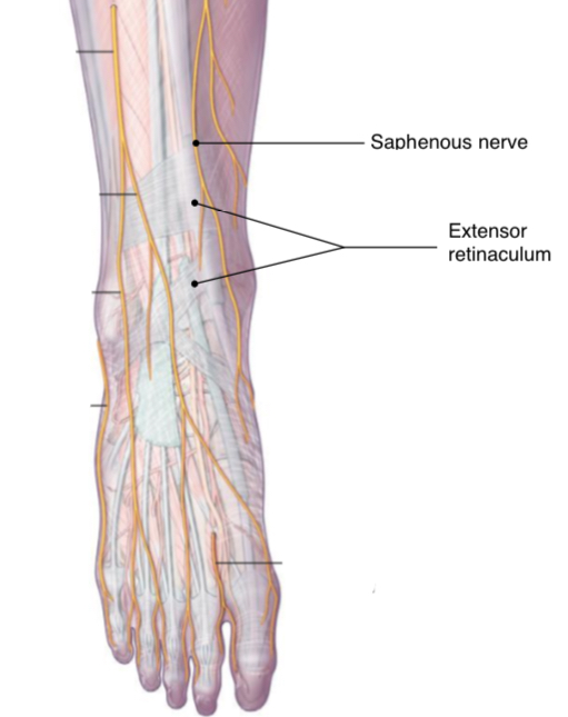
Most of the structures that pass from the leg to the foot pass beneath the extensor retinaculum, there are some structures that pass superficial(or anterior) to the extensor retinaculum.
The structure that passes superficial to the extensor retinaculum:
1. The saphenous nerve
It is a cutaneous branch of the femoral nerve . It enters the foot anterior to the medial malleolus to reach the dorsal surface. It innervates the skin over the medial aspect of the foot down to the end of the 1st metatarsal bone.
It is a cutaneous branch of the femoral nerve . It enters the foot anterior to the medial malleolus to reach the dorsal surface. It innervates the skin over the medial aspect of the foot down to the end of the 1st metatarsal bone.
Anterior view.
Right lower limb.
2. The great saphenous vein
It arises by the dorsal vein of the great toe & the venous arch at the dorsum of the foot, is ascending anterior to the medial malleolus accompanied by the saphenous nerve & continues along the anteromedial side of the leg.
It arises by the dorsal vein of the great toe & the venous arch at the dorsum of the foot, is ascending anterior to the medial malleolus accompanied by the saphenous nerve & continues along the anteromedial side of the leg.
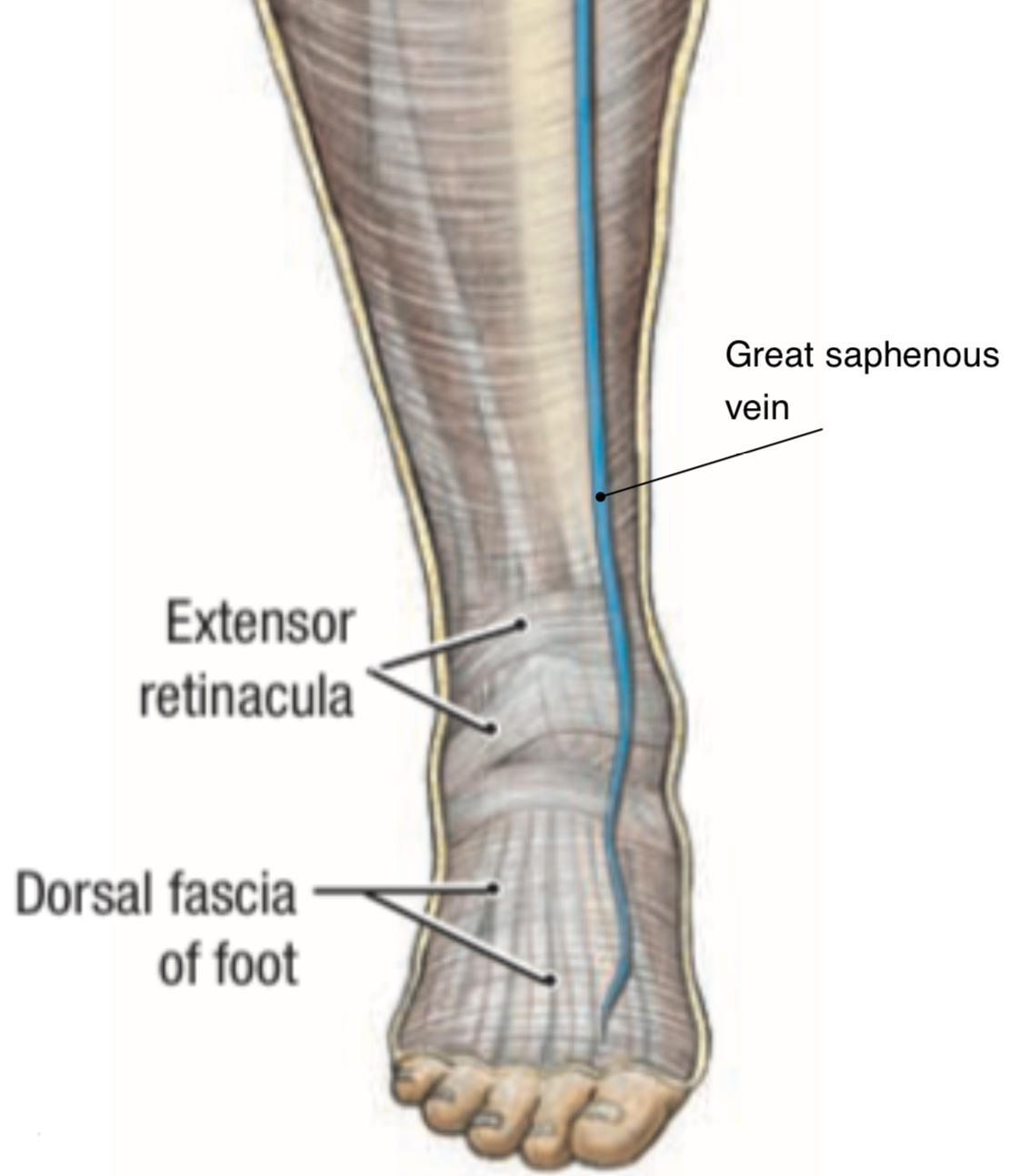
Anterior view.
Right lower limb.
3. Superficial fibular(peroneal) nerve
is one of the ultimate branches of the common fibular nerve , enters the dorsum of the foot superficial to the extensor reticulum between the fibularis brevis m. & the extensor digitorum longus m., divides into medial & lateral branches that innervate the most skin of the foot dorsum.
is one of the ultimate branches of the common fibular nerve , enters the dorsum of the foot superficial to the extensor reticulum between the fibularis brevis m. & the extensor digitorum longus m., divides into medial & lateral branches that innervate the most skin of the foot dorsum.
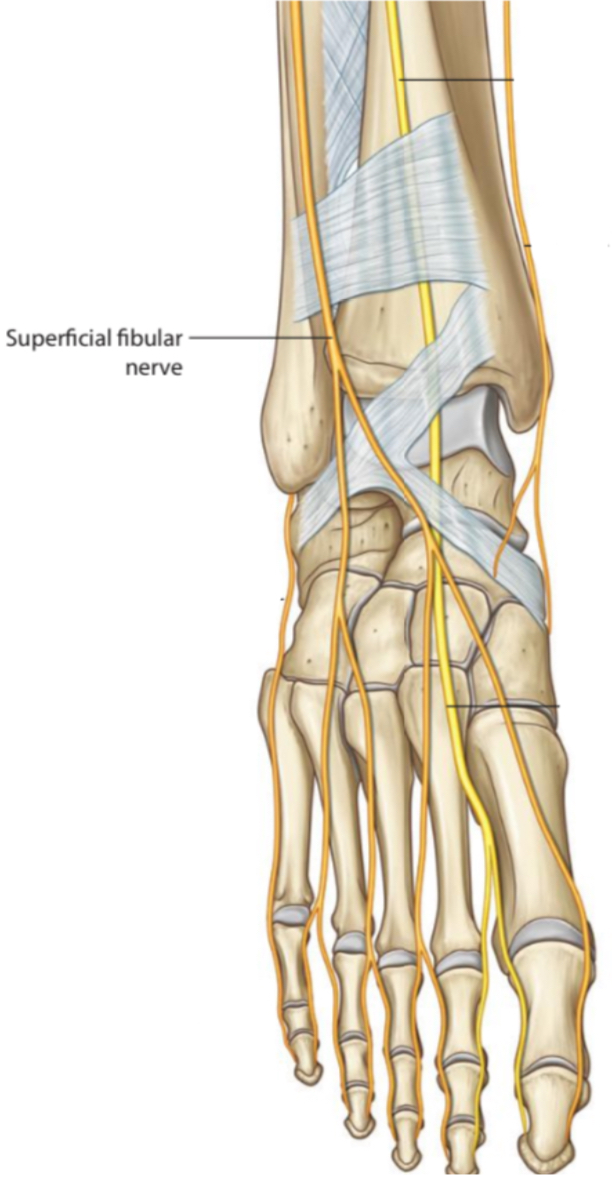
Anterior view.
Right lower limb.
Clinical Note
1. The saphenous nerve ’s important role in this region is being susceptive to damage during ankle surgery to treat bones fracture & acute arthritis, is also exposed to seizures during great saphenous vein cutdown & which leads to numbness at the medial side of the foot.
2. The great saphenous vein ’s important role i this region is being useful for some procedures like repeat blood transfusion & cannula insertion.
3. The Superficial fibular (peroneal) nerve ‘s important role in this region is entrapment due to ankle sprains & twisting that lead to the nerve extending, causing pain at the dorsal surface of the foot.
References
Moore-Clinically Oriented Anatomy (7th Edition) 535,540-541,617
Snell’s Cinical Anatomy By Regions(10th Edition) 1293-1294,1347,1353,1360,1383,1384
Saphenous Nerve - Cleveland Clinic- https://my.clevelandclinic.org/health/body/22331-saphenous-nerve
Superficial Peroneal Nerve - Chris Battista MD- Orthobullets -
https://www.orthobullets.com/anatomy/10137/superficial-peroneal- nerve
Gray’s Atlas of Anatomy (2nd Edition)-344,347,354.
Grant’s Atlas of Anatomy (13th Edition)5.15.
