Levator scapulae
By : Fahad Al-BakNaming: -
/le-VAY-tor SKAP-you-lee/
From the name we know the muscle elevate the scapula.
From the name we know the muscle elevate the scapula.
General: -
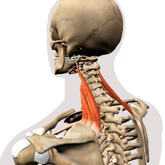
The superior third of the strap-like, levator scapulae lie deep to the sternocleidomastoid; the inferior third is deep to the trapezius. From the transverse processes of the upper cervical vertebrae, the fibers of the leva tor of the scapula pass inferiorly to the superomedial border of the scapula.

Supply: -
-Blood supply comes from The Dorsal Scapular Artery (a branch of the Subclavian Artery).
-Nerve supply comes from Dorsal Scapular Nerve C3, C4, C5.

Origin & Insertion: -
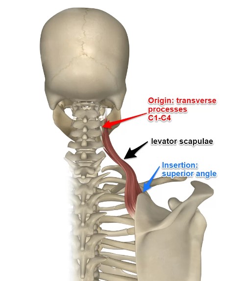
-Origin: attaches by tendinous slips to the transverse processes of the upper three or four cervical vertebrae (attaching to the posterior tubercles of the lower two) behind the attachment of scalenus medius.
-Insertion: From here the fibers run downwards and laterally to attach to the medial margin of the scapula between the superior angle and base of the spine.

Motion & Action: -
(Elevates the scapula at the Scapulocostal joint)
-Working with trapezius, levator scapulae can produce elevation and retraction of the pectoral girdle or resist its downward movement, as when carrying a load in the hand. Again, when working with trapezius, contraction of both sides produces extension of the neck, while one side produces lateral flexion of the neck. Levator scapulae also helps to stabilize the scapula and is active in producing medial rotation of the scapula.
-Working with trapezius, levator scapulae can produce elevation and retraction of the pectoral girdle or resist its downward movement, as when carrying a load in the hand. Again, when working with trapezius, contraction of both sides produces extension of the neck, while one side produces lateral flexion of the neck. Levator scapulae also helps to stabilize the scapula and is active in producing medial rotation of the scapula.
In The Field Notes: -
Levator scapulae Test: The levator scapulae length and tension can be assessed by placing the patient in supine, stabilizing the ipsilateral scapula, and contralaterally side bend and rotate the head. Also, trigger points are common in this muscle and can be palpated for in both the superior attachment and inferior attachment.
Clinical Note: -
An isolated lesion of the dorsal scapular nerve with a consequent paralysis of the levator scapulae muscle is very rare. The symptoms include “winging” of the scapula (scapula alate), as well as atrophy of both the levator scapulae and rhomboid muscles. As the affected patients may have no clear complaints, the correct diagnosis is often made too late.

Relations: -
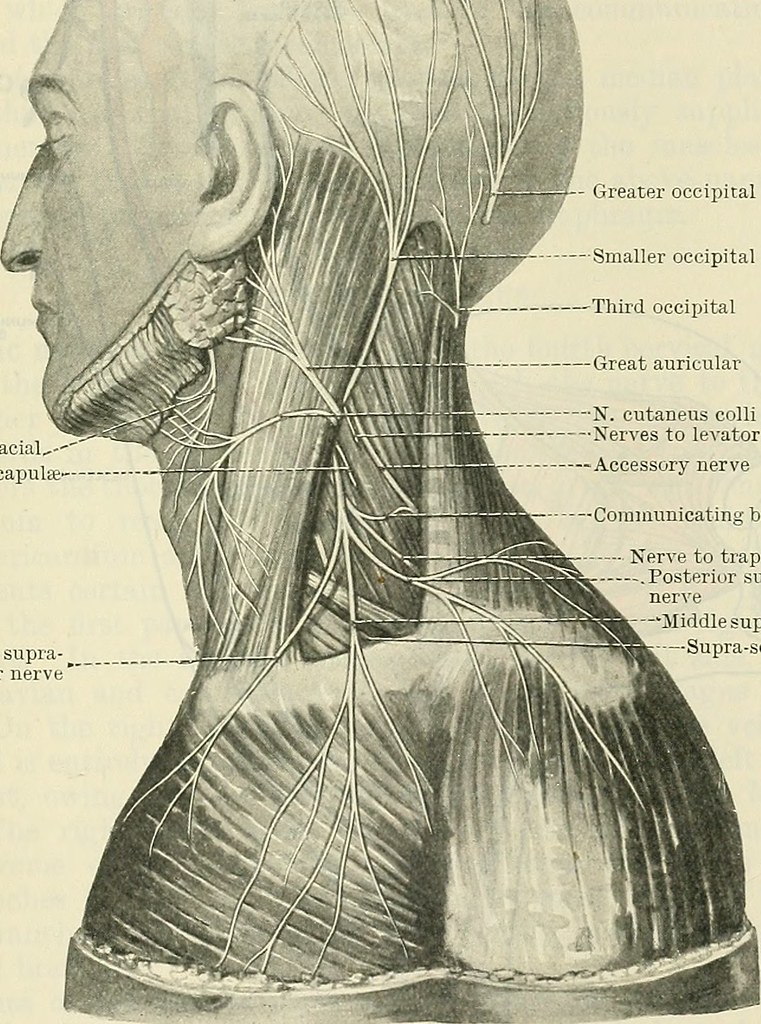
Inferiorly, the levator scapulae are deep to the trapezius. Superiorly, the levator scapulae is deep to the sternocleidomastoid.
There is a part of the middle to upper levator scapulae that is superficial between the trapezius and splenius capitis posteriorly, and the sternocleidomastoid and scalene anteriorly, in the posterior triangle of the neck.
The scapular attachment of the levator scapulae is superior to the attachment of the rhomboids.
The transverse process attachment of the levator scapulae is directly deep (anterior) to the transverse process attachment of the splenius cervicis and directly superficial (posterior) to the transverse process attachment of the scalene.
The levator scapulae are located within the deep back arm line myofascial meridian.
Sources: -
- Moore - Clinically Oriented Anatomy 7th Edition (703).
- Joseph E Muscolino - The Muscular System Manual: The Skeletal Muscles of the Human Body
Book 4t edition (102-103).
-Nigel Palastanga & Roger Soames Anatomy and Human Movement Structure and Function 6th edition (58).
- Joseph E Muscolino - The Muscular System Manual: The Skeletal Muscles of the Human Body
Book 4t edition (102-103).
-Nigel Palastanga & Roger Soames Anatomy and Human Movement Structure and Function 6th edition (58).
