


carpal Tunnel
By : Ruqayah AkramDefinition
It is a passageway found on anterior wrist for structures passage between anterior forearm and hand.
Boundaries
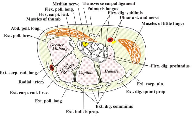
The floor: carpal groove or (carpal arch) a deep arch formed by the palmar aspect of the carpal bones.
This arch (carpal arch) is bounded :
Medially : the pisiform bone and the hook of the hamate.
Laterally: the tubercles of the scaphoid and trapezium bones.
The roof: flexor retinaculum (also known as transverse carpal ligament).
note:
This arch (carpal arch) is bounded :
Medially : the pisiform bone and the hook of the hamate.
Laterally: the tubercles of the scaphoid and trapezium bones.
The roof: flexor retinaculum
Note
Flexor retinaculum: a thick connective tissue ligament.note:
Note
This ligament bridges the space between the medial and lateral ends of the carpal arch, converting the arch into a tunnel.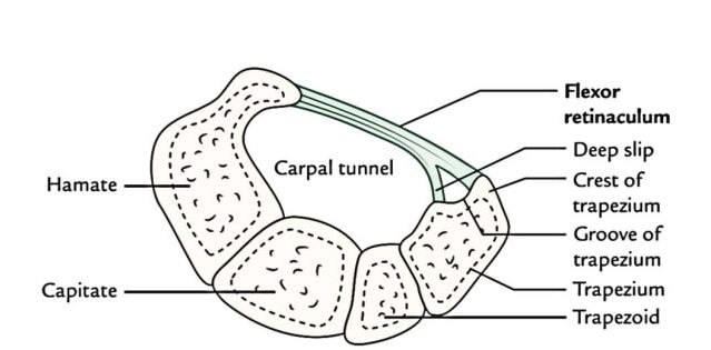
.jpg)
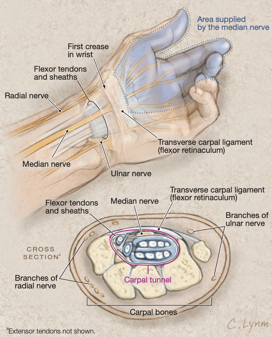
Carpal Tunnel Contents
The carpal tunnel contains
1.Median nerve
2.nine tendons, which they are:
a- flexor digitorum superficialis(four tendons)
b- flexor digitorum profundus(four tendons)
c- flexor pollicis longus (one tendon)
1.Median nerve
2.nine tendons, which they are:
a- flexor digitorum superficialis(four tendons)
b- flexor digitorum profundus(four tendons)
c- flexor pollicis longus (one tendon)
content of carpal tunnel
Notes:
1.The tendons are enveloped by synovial tendinous sheaths that allow free movement between them.
2.The eight tendons of both flexor digitorum profundus and flexor digitorum superficialis are surrounded by a single synovial sheath, otherwise the flexor pollicis longus’s single tendon is surrounded by its own synovial sheath.
3.All these tendons are hold in place by the flexor retinaculum
2.The eight tendons of both flexor digitorum profundus and flexor digitorum superficialis are surrounded by a single synovial sheath, otherwise the flexor pollicis longus’s single tendon is surrounded by its own synovial sheath.
3.All these tendons are hold in place by the flexor retinaculum
carpal tunnel syndrome
Definition
Carpal tunnel syndrome refers to a group of signs and symptoms that result from the compression of the median nerve as it travels through the carpal tunnel.
The Reasons
Any agent narrows the tunnel can cause the carpal tunnel syndrome, as :
1.wrist fracture
2. swelling of the long tendons that pass through the carpal tunnel.
Note:
Risk factors for this condition include:
1.diabetes
2. obesity
3. pregnancy
4.arthritis
5. trauma
6. Occupational biomechanical factors as :
1)repeated and forceful movements of the hand and wrist
2) use of hand-held powered vibratory tools.
The Symptoms
1.Pain
2.Paresthesia
3. Less commonly ,weakness in the median nerve distribution, which innervates:
a. Sensory innervation to the skin of the palmar side of the thumb, index finger, middle finger and half of the ring finger.
b. Motor innervation to the thenar eminence muscles, These muscles start to atrophy and disappear during carpal tunnel syndrome.
The Treatment
Options include
1.the use of splints
2.corticosteroid injections
3. some exercise.
As the case severity increases, the surgical decompression is needed.
1.the use of splints
2.corticosteroid injections
3. some exercise.
As the case severity increases, the surgical decompression is needed.
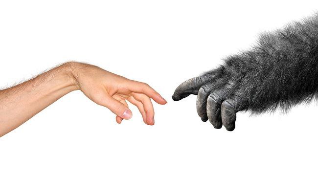
Ape like hand
carpal tunnel syndrome can causes Ape like hand
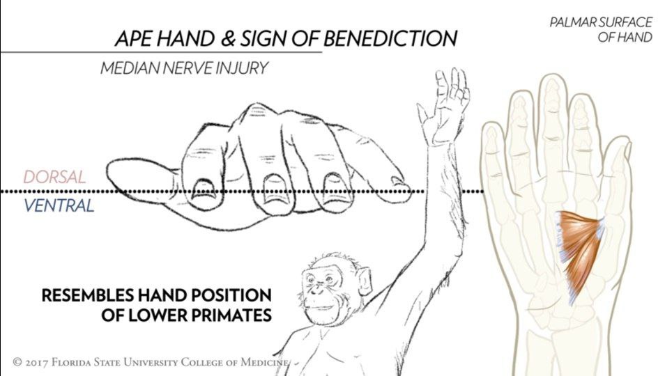
1-In the normal hand at rest, digits 2 to 5 are loosely flexed and all are on the same dorsal ventral plane.
The thumb is held in a more ventral plane due primarily to the tonic action of the thenar muscles (abductor pollicis brevis , and opponens pollicis )
The thumb is held in a more ventral plane due primarily to the tonic action of the thenar muscles (abductor pollicis brevis , and opponens pollicis )
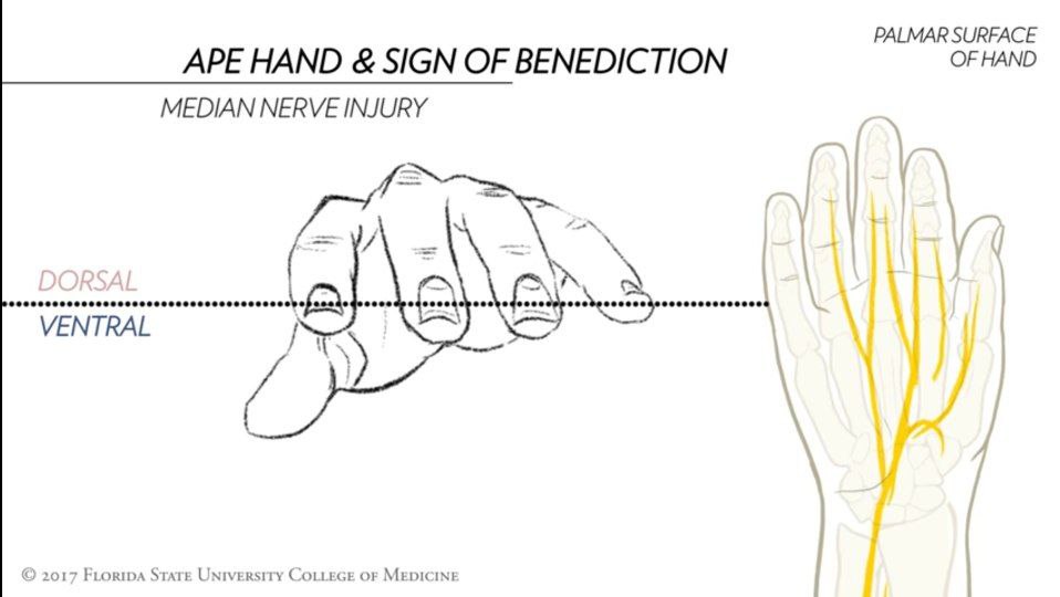
2- Following the median nerve injury, the thenar muscles are paralyzed and the thumb is pulled into the plane of the other digit by unopposed action of adductor pollicis muscle .
The position of the hand with all of the digits aligned in the same dorsal ventral plane resembles the hand position primates hence the term ‘’APE HAND’’
The position of the hand with all of the digits aligned in the same dorsal ventral plane resembles the hand position primates hence the term ‘’APE HAND’’
References
1.Keith L. Moore , Arthur F. Dalley A. M. R. Agur; Moore clinically oriented anatomy 7thedition;pp.689,746,750,767-768,773,786,789-791,817
2.Dr. Lawrence E. Wineski,PhD;SNELLIS CLINICAL ANATOMY BY REGIONS 10th edition;pp.234-235,285,291,293-294,297,346,360-361,466
3.MRI of the wrist: normal anatomy; Adrian Rad,BSc;Kenhub; https://www.kenhub.com/en/library/anatomy/wrist-mri
4.Carpal tunnel; Sara Ferreira,MD;Kenhub; https://www.kenhub.com/en/library/anatomy/carpal-tunnel
5.Median Nerve Palsy; Donald D. Davis , Steven M. Kane; National Library of Medicine;https://pubmed.ncbi.nlm.nih.gov/32491813/
references of image
cover image: https://www.google.com/url?sa=i&url=https%3A%2F%2Forthoinfo.aaos.org%2Fen%2Fdiseases--conditions%2Fcarpal-tunnel-syndrome%2F&psig=AOvVaw3GatHqYVs57y6GXsU5Abgx&ust=1664043011683000&source=images&cd=vfe&ved=0CAwQjRxqFwoTCJj9gbrBq_oCFQAAAAAdAAAAABAD
fig-1,2:boundaries of carpal tunnel
https://www.google.com/url?sa=i&url=https%3A%2F%2Fwww.earthslab.com%2Fanatomy%2Fcarpal-tunnel%2F&psig=AOvVaw2zvtBnr_OxvFAitMMVzr2K&ust=1663887624820000&source=images&cd=vfe&ved=0CAwQjRxqFwoTCPCmu9P-pvoCFQAAAAAdAAAAABAM
fig3: content of carpal tunnel:https://www.google.com/url?sa=i&url=https%3A%2F%2Fbrownmedpedsresidency.org%2Febpe-cts%2F&psig=AOvVaw1-nQmxmVe7cLV6DlEXhVKx&ust=1664036554648000&source=images&cd=vfe&ved=0CAkQjRxqFwoTCNjLurapq_oCFQAAAAAdAAAAABAM
fig4 :https://www.google.com/imgres?imgurl=https%3A%2F%2Flivewellcc.com%2Fwp-content%2Fuploads%2F2020%2F01%2FWhat-Is-Carpal-Tunnel-Syndrome-and-How-Can-You-Treat-It-.png&imgrefurl=https%3A%2F%2Flivewellcc.com%2Fwhat-is-carpal-tunnel-syndrome-and-how-can-you-treat-it%2F&tbnid=7Nbxcxp0CO4OPM&vet=12ahUKEwjlqdLZhaf6AhWMNewKHZBtCyEQMyg4egQIARAz..i&docid=AFDXcljL4x1t9M&w=845&h=355&itg=1&q=carpal%20tunnel&hl=ar&ved=2ahUKEwjlqdLZhaf6AhWMNewKHZBtCyEQMyg4egQIARAz
