Distal Radioulnar Joint
By : Yaser MohammedLocation and Articulation
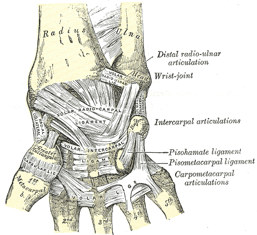
Distal radioulnar joint: is a joint in the upper limb, especially in the forearm proximal to the wrist joint lying between the distal heads of radius and ulna bones.
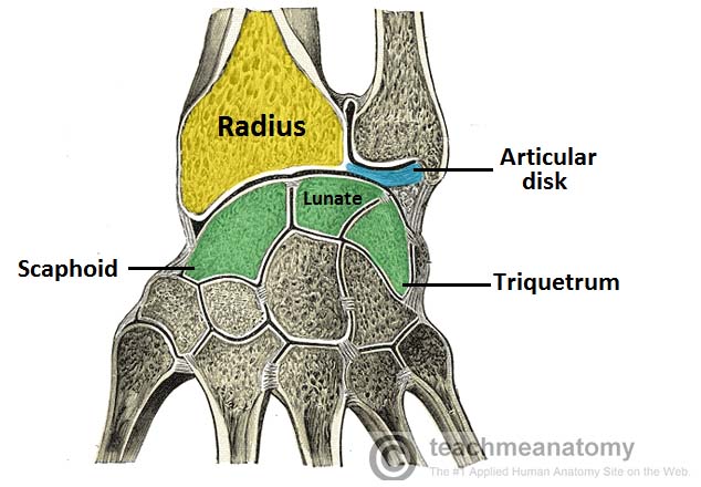
The distal radioulnar joint formed from the articulation between the distal head of ulna (crescent and convex shaped) and the ulnar notch of radius (concave shaped), (the ulnar head sits within the sigmoid notch of the radius), Both surfaces are lined by the hyaline cartilage. [see fig 1]
The distal radioulnar joint formed from the articulation between the distal head of ulna (crescent and convex shaped) and the ulnar notch of radius (concave shaped), (the ulnar head sits within the sigmoid notch of the radius), Both surfaces are lined by the hyaline cartilage. [see fig 1]
Bones Articulation in the Distal Radioulnar Joint
Articular Disk
This joint contains a triangular fibrocartilaginous ligament, called the Articular disk . The apex of the disc is attached to the styloid process of ulna, while the base is anchored to the ulnar notch of radius. It serves two functions :
• Connects the radius and ulna together and holds them together during movement at the joint.
• Separates the distal radioulnar joint from the wrist joint.
• Connects the radius and ulna together and holds them together during movement at the joint.
• Separates the distal radioulnar joint from the wrist joint.
Joint Type and Its Movement
Like the proximal radioulnar joint this joint is a synovial pivot joint, uniaxial (move in only one plane), it is a critical stabilizing structure of the ring stabilizing the radius and ulna,
as it is a pivot joint so it moves in only one degree of motion it works with the proximal radioulnar joint to do these two actions:
Pronation: (range of motion is 61–66° ) When the palm or forearm faces down, it's pronated. the muscles that work to form pronation at this joint are the pronator quadratus and pronator teres . The first is for slight movements, while the second is included in fast movements and movements against resistance.
Supination: (range of motion is 70–77° ) When the palm or forearm faces up, it's supinated. when the forearm is extended, supination is formed by contraction of the supinator muscle , while when it's extended, and for the movement against resistance, the biceps brachii muscle acts as an accessory supinator.[see fig 3]
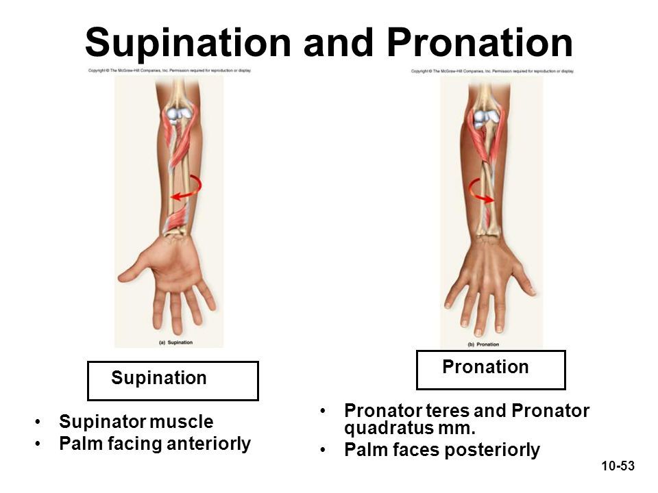
Movements of this joint
Blood Supply
The Blood supply of distal radioulnar joint is the palmar and dorsal branches of the anterior interosseous artery. [see fig 4,5]
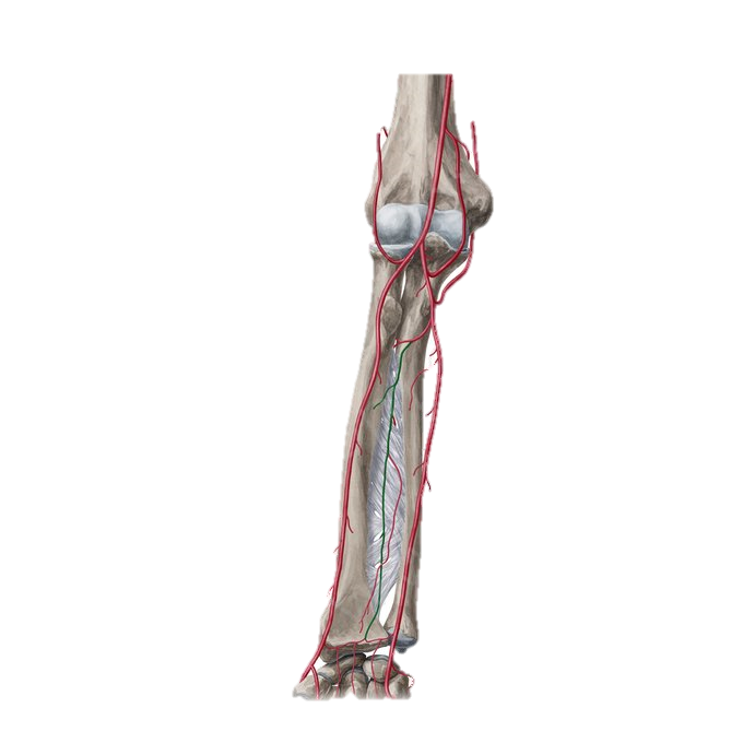
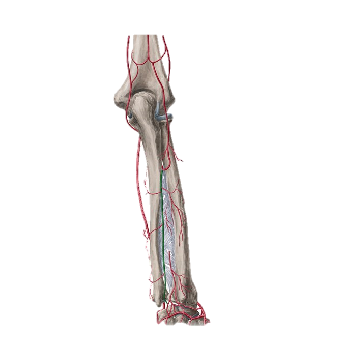
Blood Supply of Distal Radioulnar Joint
Nerve Innervation
while the Nerve innervation of it comes from the branches of the anterior and posterior interosseous nerves. The first is a branch of the median nerve, while the second stems from the radial nerve.
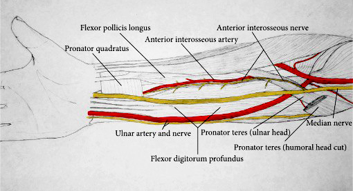
The Nerve innervation of the joint
Capsule and Ligaments
The joint is enclosed by a fibrous capsule that attaches to the margins of the articular surfaces.
There are many structures that work together to provide stability to this joint we can characterize them as extrinsic and intrinsic stabilizers. the extrinsic stabilizers are the tendons of extensor carpi ulnaris, pronator quadratus (These two cross the joint and hold it tightly) and the interosseous membrane of the forearm. While the intrinsic stabilizers are the joint capsule, triangular fibrocartilage complex (TFCC), and distal radioulnar ligaments.
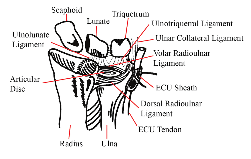
The Triangular Fibrocartilage Complex (TFCC) is a biconcave ligamentous complex that stabilizes and cushions the joints of the wrist region (which are; distal radioulnar, ulnocarpal, and radiocarpal joints). It consists of the articular disc of the distal radioulnar joint, ulnar collateral ligament, dorsal and palmar radioulnar ligaments, the base of the extensor carpi ulnaris sheath, and the ulnolunate and ulnotriquetral ligaments.
The core of the TFCC is the articular disc of the distal radioulnar joint. The dorsal and palmar parts of the TFCC are thickened and known as the dorsal and palmar radioulnar ligaments, respectively. Each of these ligaments consists of superficial and deep components which differ by their ulnar attachments. The superficial components insert onto the styloid process of ulna, while the deep ones insert slightly more laterally. The ulnar collateral, ulnolunate and ulnotriquetral ligaments join the TFCC on its ulnar attachment. The dorsal margin of the TFCC is fused with the floor of the base of the extensor carpi ulnaris sheath.
The Function of the TFCC is to stabilize the joints within the wrist region by transmitting and distributing the load from the hand to the ulna. [see fig 7]
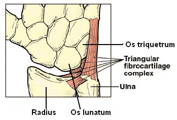
The Triangular Fibrocartilage Complex (TFCC)
