


Hip Joint
By : Yaser MohammedLocation and Articulation
Hip Joint: Is a joint in the lower limp, it works as a connection area between the axial skeleton and the lower limp.
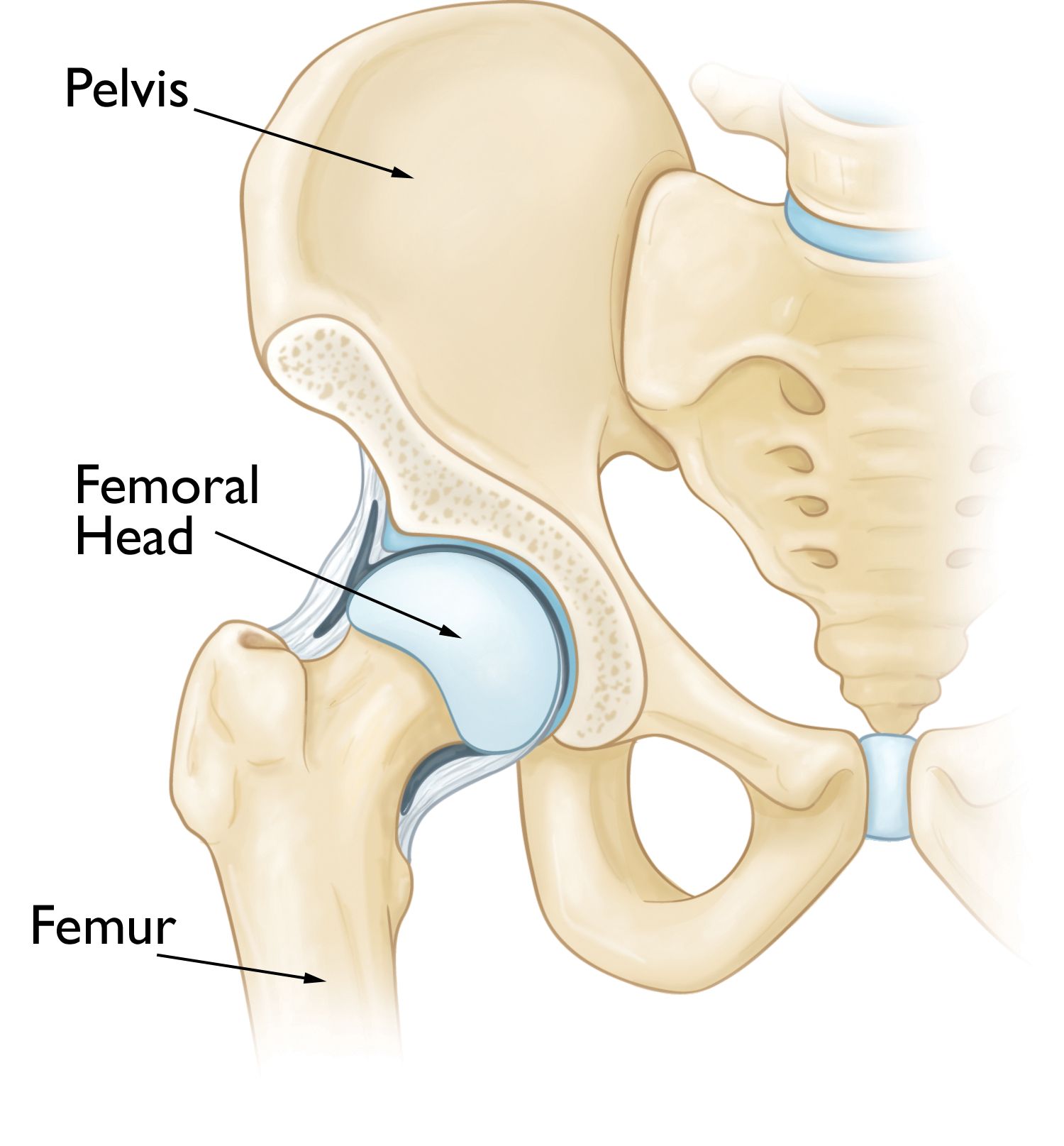
The hip joint is formed from the articulation between the proximal ending of the femur and the inferolateral aspect of the hip bone , the femur head fits into the concavity of the acetabulum to make this joint. The ellipsoid head of the femur is covered with hyaline cartilage except for a rough central depression, the fovea capitis .
On the other hand, the hip bone is actually formed from three bones which are: the ilium, ischium, and pubic bones . and the acetabulum which articulates within the hip joint formed from the fusion of these bones.
This structure of acetabulum gives the joint high stability making it the most stable joint in the body, that's very important because all body weight (during standing) is transferred to the lower limp through this joint.


Hip Joint Articulation
Joint Type and Its Movement
The Joint is a Ball-and-Socket Synovial joint, multiaxial (moves in three degrees of freedom) the movements of the joint are: flexion-extension, abduction- adduction, external rotation- internal rotation, and circumduction.
Flexion: The range of movement ROM of this action is 120 degrees, other says 100 degrees, The flexion of the hip joint draws the thigh towards the trunk(moving thigh anteriorly), main flexors muscles of the joint are the iliopsoas muscle (psoas major and iliacus) and the rectus femoris muscle, and are assisted by the pectineus, tensor fasciae latae, and sartorius muscles.
Extension: Range of motion is 0 - 115 degrees. Opposite flexion, moving the thigh away from the trunk(moving thigh posteriorly), The primary extensor of the joint is the gluteus maximus muscle , assisted by the hamstring muscles (biceps femoris, semitendinosus, semimembranosus) and the adductor magnus muscle.
Abduction: Range of motion is 0 to 45 degrees. Abduction of the hip is the movement of the leg away from the midline, The primary abductors of the hip joint are the gluteus medius and the gluteus minimus muscles. The tensor fasciae latae, piriformis, and sartorius muscles also assist in hip abduction.
Adduction: The range of motion, same as abduction, is 0 to 45 degrees. Adduction of the hip is the movement of the leg closer to the midline, The adductors of the hip joint are the adductors muscles : longus, brevis, and magnus, and the gracilis muscle, assisted by pectineus, quadratus femoris, and the inferior part of gluteus maximus.
The internal and external rotation of the hip joint both occurs in the horizontal plane at a range of motion of about 45 degrees
Internal Rotation (medial rotation): The internal rotation is moving the thigh bone inward, it acts by the anterior fibers of gluteus medius and minimus muscles , and is also assisted by tensor fascia latae.
External Rotation (lateral rotation): The external rotation is when the thigh and knee rotate outward, away from the body, it is produced by biceps femoris, gluteus maximus, and piriformis and is assisted by the obturators, Gemelli, and quadratus femoris.


Hip joint movements
Blood Supply
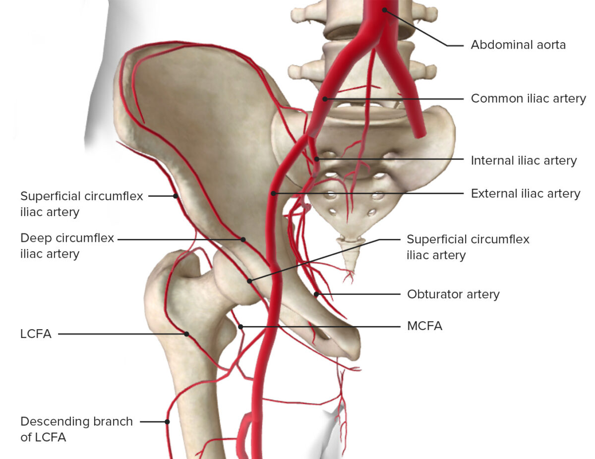
The Blood supply of the hip joint comes from anastomoses formed by the medial and lateral circumflex femoral arteries, which are branches of the profunda femoris artery (deep femoral artery), the obturator artery, and the superior and inferior gluteal arteries.

The Blood supply of Hip joint
Nerve Innervation
While the Nerve Innervation of the joint comes from nerves that emerge from the lumbosacral plexus (L2-S1) as the following:
-The anterior aspect is innervated by the femoral nerve.
-The inferior aspect is supplied by the obturator nerve.
-The superior aspect is innervated by the superior gluteal nerve
-While the nerve to the quadratus femoris innervates the posterior aspect .
-The anterior aspect is innervated by the femoral nerve.
-The inferior aspect is supplied by the obturator nerve.
-The superior aspect is innervated by the superior gluteal nerve
-While the nerve to the quadratus femoris innervates the posterior aspect .
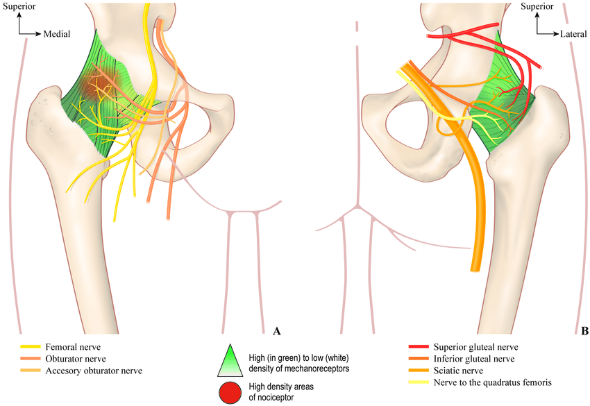
The sensory innervation of the hip joint: (A) anterior, and (B) posterior views.
Note: These nerves are the same nerves that innervate the knee joint, which explains why pain can be referred to the knee from the hip and vice versa.
Joints Capsule and Ligaments
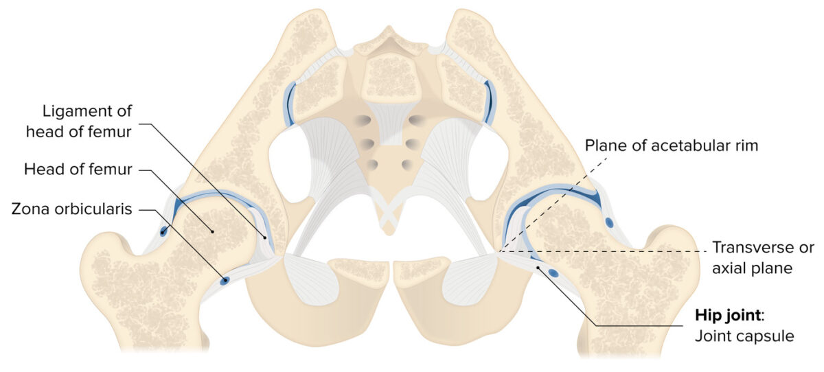
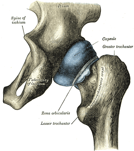
The hip joint is enclosed by a dense and strong fibrous capsule , this capsule is attached proximally by the acetabular labrum and close to the transverse acetabular ligament.
on the other side, it's attached to the femur at the intertrochanteric line anteriorly, and about 4 cm medial to the intertrochanteric crest( which means that only half of the neck is enclosed).
The hip joint has many strong ligaments that play important role in stabilizing it through movement, these ligaments are classified into Extracapsular and Intracapsular ligaments.
The Capsule of hip joint
The Extracapsular Ligaments
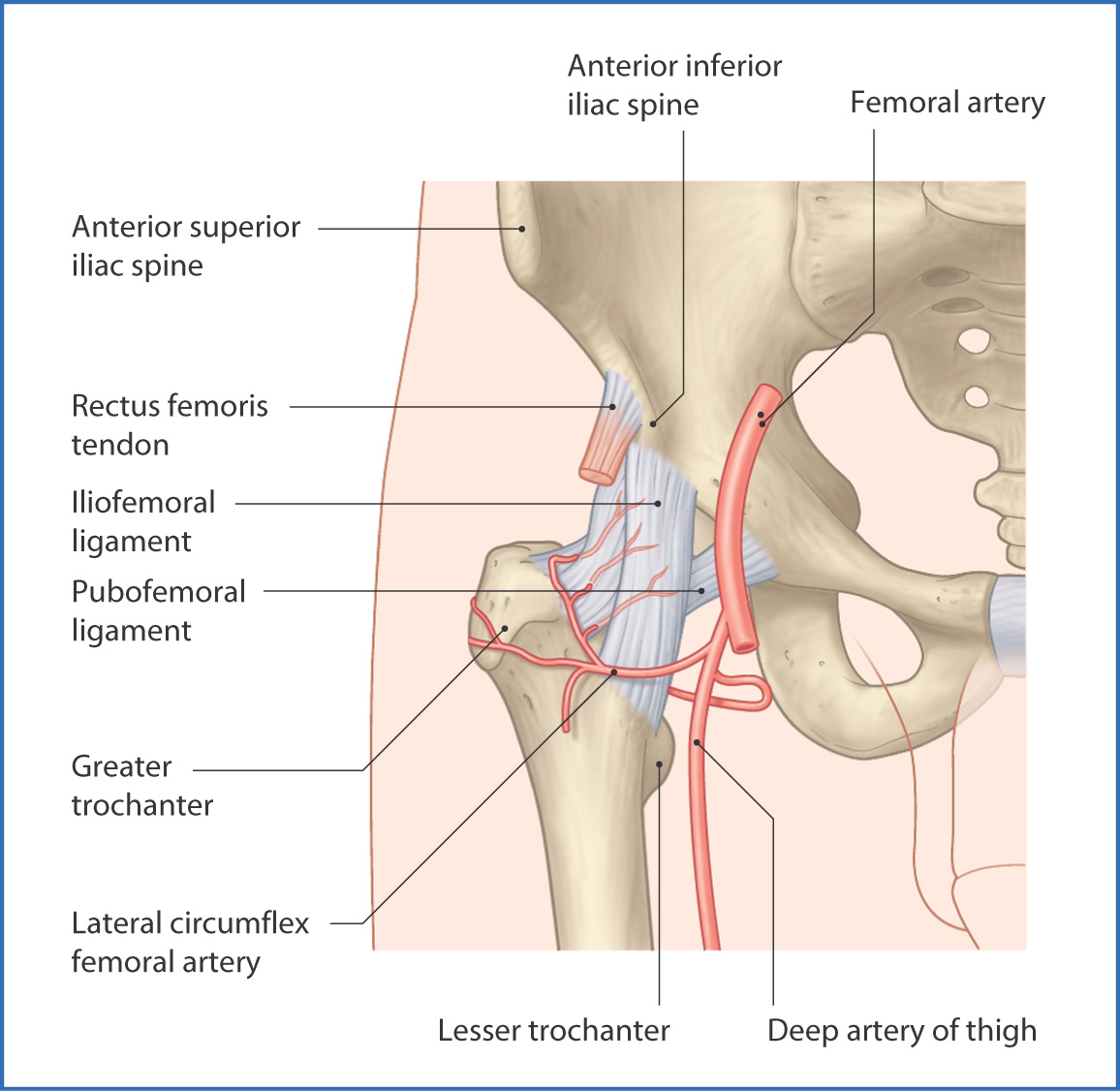
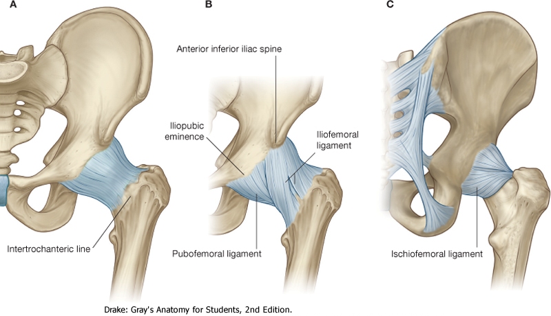
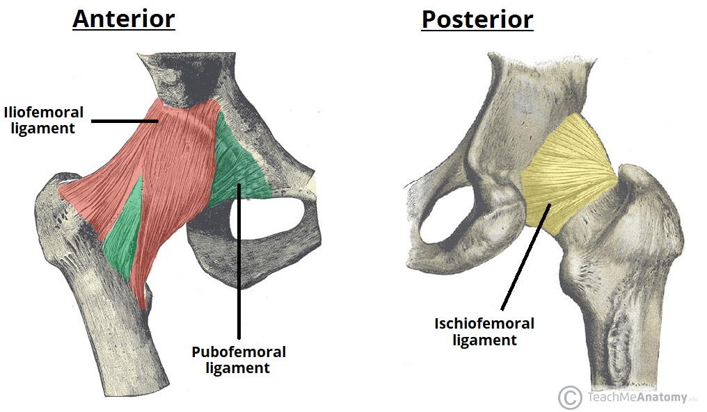
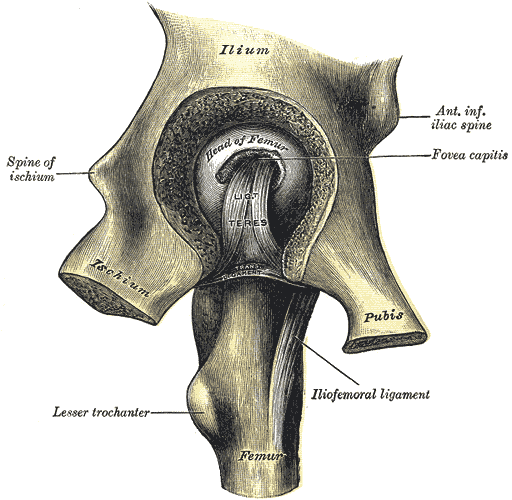
Iliofemoral ligament: is the strongest ligament in the body, it is a triangular Y-shaped ligament, that extends proximally from the anterior inferior iliac spine and continues until it attaches to the intertrochanteric line of the femur distally. its function is to prevent hyperextension of the hip joint when standing.
Pubofemoral ligament: it also has a triangular shape, that extends from the superior pubic rami to the intertrochanteric line of the femur.
Its function is to reinforce the joint capsule (anteriorly and inferiorly) and prevent excessive abduction and extension.
Ischiofemoral: is the weakest of the other three ligaments, lies posteriorly, it extends from the body of the ischium to the greater trochanter of the femur.
Its function is to reinforce the joint capsule (posteriorly) and prevents hyperextension and holds the femoral head in the acetabulum.
Hip anatomy: The Ligaments of the Hip joints
The Intracapsular Ligament
Ligament of head of femur: its the only intracapsular ligament in the hip joint, which is a flattened triangular band of connective tissue which extends from the acetabular fossa to the fovea capitis of the femur.
-it has no function in the stability of the joint
It carries a small branch of the obturator artery, which is the artery to the head of the femur one of the blood suppliers of the hip joint.
-it has no function in the stability of the joint
It carries a small branch of the obturator artery, which is the artery to the head of the femur one of the blood suppliers of the hip joint.
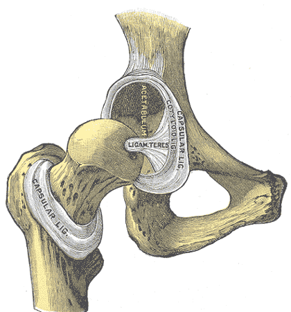
The Intracapsular ligament:
Ligament of head of femur
References
1) Moore K. L., Dalley, A. F., & Agur, A. M. R. (2014)- Clinically Oriented Anatomy 7th Edition (626-634 plates).
2) Netter, F. (2019). Atlas of Human Anatomy (7th ed.). Philadelphia, PA: Saunders.
3) Hip joint, Edwin Ocran MBChB, MSc, Kenhub.
https://www.kenhub.com/en/library/anatomy/hip-joint
4) The Hip Joint, Oliver Jones , Teachne Anatomy.
https://teachmeanatomy.info/lower-limb/joints/hip-joint/
5) Hip Anatomy, Physiopedia.
https://www.physio-pedia.com/Hip_Anatomy
References of images
Cover Image: Cross-section of the hip joint, featuring the insertions of the articular capsule and supporting ligaments, Lecturio.
https://cdn.lecturio.com/assets/Cross-section-of-the-hip-joint-scr-1200x542.jpg
fig 1: American Academy of Orthopaedic Surgeons Osteonecrosis of the Hip - OrthoInfo - AAOS. https://orthoinfo.aaos.org/link/c3372cd573fe45ad8225ceeb9d83535b.aspx
fig 2: Lateral view of the acetabulum and hemipelvis. Musculoskeletal Key.
https://i2.wp.com/musculoskeletalkey.com/wp-content/uploads/2020/06/10-1055-b-003-121618_c002_f001-2.jpg?w=960
fig 3: The three bones which meet to form the acetabulum, The university of Liverpool.
https://www.liverpool.ac.uk/~trh/local_html/hipfracture/images/acetabulum.jpg
fig 4: Lower Limb | Basicmedical Key. https://basicmedicalkey.com/wp-content/uploads/2016/06/B9781455710782000067_f006-003-9781455710782.jpg
fig 5: Anatomy 101 - The hips — YOGARU. https://images.squarespace-cdn.com/content/v1/55a9457de4b0e82a7749b6d3/71c71e9d-dccc-49e9-a95b-47936b0bc523/PAUSE_0164_C.png
fig 6: Hip Anatomy - Recon - Orthobullets. https://upload.orthobullets.com/topic/12769/images/f041-001-9780323077798.jpg
fig 7: Blood supply of hip joint, Lecturio. https://cdn.lecturio.com/assets/Blood-supply-of-the-hip-joint-scr.png
fig 8: Blood supply of hip joint, Lecturio. https://cdn.lecturio.com/assets/Blood-supply-of-the-hip-joint-1200x926.jpg
fig 9: Summary of the sensory innervation of the hip joint based on review of the literature: (A) anterior, and (B) posterior views. Pierre Laumonerie , ReasearchGate.
https://www.researchgate.net/profile/Pierre-Laumonerie/publication/349216762/figure/fig2/AS:990083582787586@1613065729640/Summary-of-the-sensory-innervation-of-the-hip-joint-based-on-review-of-the-literature.png
fig 10: Henry Gray (1918) Anatomy of the Human Body, plate 343
fig 11: The Ligaments of the Hip joints, How To relief. https://www.howtorelief.com/wp-content/uploads/2017/09/Ligaments-of-the-Hip-joints.jpg
fig 12: The extracapsular ligaments of the hip joint; ileofemoral, pubofemoral and ischiofemoral ligaments. TeachMeSeries Ltd (2022). https://teachmeanatomy.info/wp-content/uploads/Extracapsular-Ligaments-of-the-Hip-Joint.-1.jpg
fig 13: Henry Gray (1918) Anatomy of the Human Body , plate 342.
fig 14: Henry Gray (1918) Anatomy of the Human Body , plate 341.
